Latin: glandula suprarenalis
Overview
- Paired endocrine gland on the upper pole of each kidney; also called the adrenal gland.
- Lies inside the renal fascia but is separated from the kidney by fat, so it stays in place when the kidney moves.
- Weighs about 4–5 g; the right gland is pyramidal, the left is crescent-shaped.
- Consists of two different tissues: an outer cortex making steroid hormones and an inner medulla making adrenaline and noradrenaline.
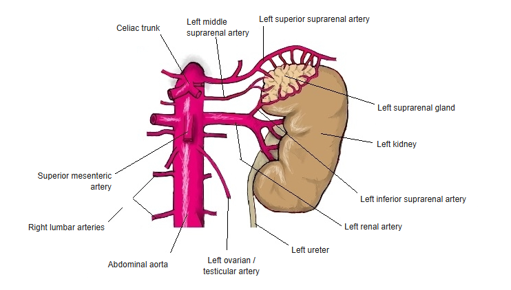
Left kidney, suprarenal gland and ureter
Location and relations
- Retroperitoneal, at about the level of T11–T12, against the crura of the diaphragm.
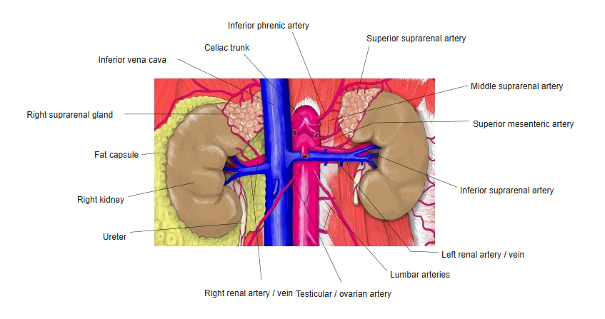
- Right gland: behind the inferior vena cava and the bare area of the liver.
- Left gland: on the medial border of the left kidney above the hilum; in front lie the stomach (through the omental bursa), pancreas and splenic vessels.
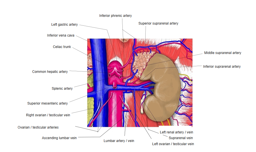
Kidneys, suprarenal glands and their arteries
Structure and hormones
| Part | Zone | Main hormones |
|---|---|---|
| Cortex | Zona glomerulosa (outer) | Mineralocorticoids – aldosterone (sodium and water retention, blood pressure) |
| Zona fasciculata (middle) | Glucocorticoids – cortisol (glucose metabolism, stress response, anti-inflammatory) | |
| Zona reticularis (inner) | Adrenal androgens (DHEA) | |
| Medulla | Chromaffin cells | Adrenaline and noradrenaline ("fight or flight") |
- The cortex develops from mesoderm; the medulla from neural crest cells and acts like a modified sympathetic ganglion.
- Cortisol and adrenal androgens are controlled by ACTH from the pituitary gland; aldosterone mainly by the renin–angiotensin system and potassium.
Blood supply and nerves
| Artery | Origin |
|---|---|
| Superior suprarenal arteries | Inferior phrenic artery |
| Middle suprarenal artery | Abdominal aorta |
| Inferior suprarenal artery | Renal artery |
- Veins: a single suprarenal vein on each side – the right drains into the inferior vena cava, the left into the left renal vein.
- Nerves: preganglionic sympathetic fibres (mainly from the splanchnic nerves via the celiac plexus) end directly on the medullary chromaffin cells.
Arteries and veins of the left suprarenal gland
References and sources
- Further reading: Suprarenal gland, Wikipedia.