Anal columns (Latin: columnae anales)
Also called the rectal columns or the columns of Morgagni
- Overview
- Reference websites
- (Wikipedia, websites with illustrations)
- Where it is found
- Related terms
Overview
· Also called the columns of Morgagni or rectal columns.
of the
anal canal
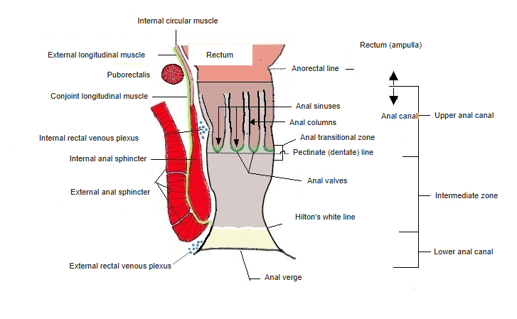
- Upper anal canal
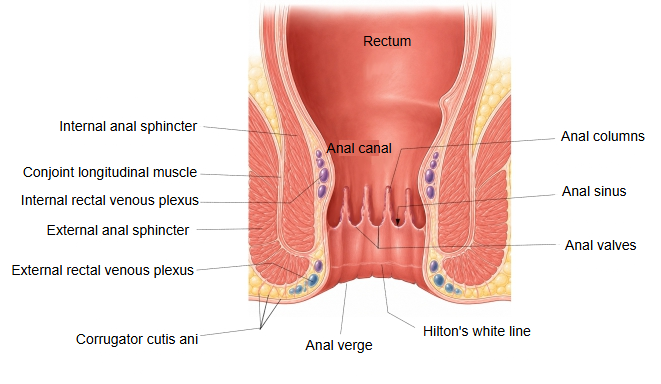
(anal column zone) : from the anorectal line to the pectinate line; about 1.5–2 cm. Lined by simple columnar epithelium continuous with the rectal mucosa. Here are the anal columns, anal sinuses and anal valves. - Intermediate zone
(transitional zone) : from the pectinate line to Hilton's white line; about 1–1.5 cm. The lining changes to stratified squamous epithelium; in places columnar and stratified squamous epithelium lie side by side. A venous plexus is well developed beneath the mucosa. - Lower anal canal
(skin zone) : from Hilton's white line to the anal verge; a few millimetres to 1 cm. Covered by stratified squamous epithelium rich in melanin pigment. It ends at the anus, whose margin is the anal verge; sweat glands are found here.
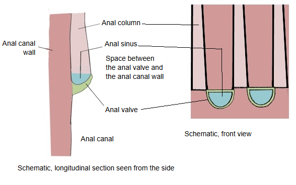
· At the bases of the anal columns the mucosa projects into the lumen as the anal valves, which join the lower ends of neighbouring columns; between each valve and the wall of the canal is a small pocket open upward, the anal sinus (anal crypt).
· The columns are mainly longitudinal ridges of the mucosa and the tissue beneath it.
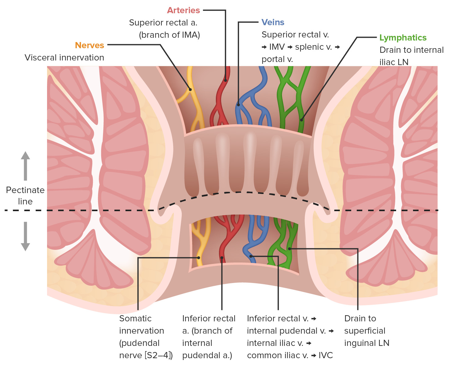
· They are raised mainly by the rich venous plexus under the mucosa and by bundles of longitudinal smooth muscle. Each column contains terminal branches of the superior rectal artery and vein; the veins form part of the internal rectal venous plexus, and enlargement of these vessels gives internal haemorrhoids.
· The columns are most obvious when the canal is empty and flatten out when it is stretched.
Reference websites
[Wikipedia]
· Anal canal, Wikipedia.
· Pectinate line, Wikipedia.
[Websites with illustrations]
· Anus and rectum (textbook figure) (Dentistry Key)

· Anal canal (book figure) (Springer Nature)

· Anal canal anatomy (journal figure) (Elsevier)

· Anal neurovasculature above and below the pectinate line (Lecturio)
Where it is found
Related terms
- Anal valves
- Anal glands
- Anorectal line
- Hilton's white line
- Anal canal
- Internal rectal venous plexus
- Rectum
References and sources
- Further reading: Anal canal, Wikipedia.
- Further reading: Pectinate line, Wikipedia.
- Federative International Programme for Anatomical Terminology (FIPAT). Terminologia Anatomica, 2nd ed. (TA2), 2019.