Pectinate line (Latin: linea pectinata)
Also called the dentate line
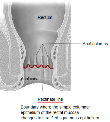
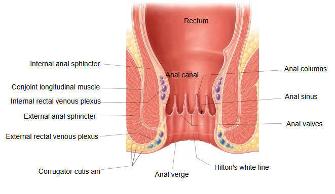
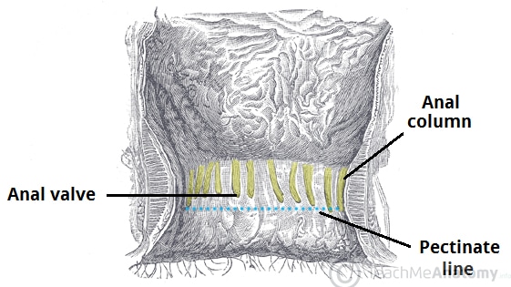
· It is formed by the row of anal valves at the lower ends of the anal columns, which gives it its toothed (comb-like) outline. It lies about 2 cm above the anal verge and is an important landmark in surgery of the anal canal.
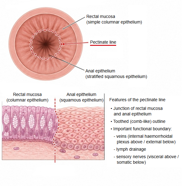
· It marks the embryological junction between the hindgut (endoderm) and the proctodeum (ectoderm), so the blood supply, venous and lymph drainage and nerve supply differ above and below it.
of the
anal canal
- Upper anal canal
(anal column zone) : from the anorectal line to the pectinate line; about 1.5–2 cm. Lined by simple columnar epithelium continuous with the rectal mucosa. Here are the anal columns, anal sinuses and anal valves. - Intermediate zone
(transitional zone) : from the pectinate line to Hilton's white line; about 1–1.5 cm. The lining changes to stratified squamous epithelium; in places columnar and stratified squamous epithelium lie side by side. A venous plexus is well developed beneath the mucosa. - Lower anal canal
(skin zone) : from Hilton's white line to the anal verge; a few millimetres to 1 cm. Covered by stratified squamous epithelium rich in melanin pigment. It ends at the anus, whose margin is the anal verge; sweat glands are found here.
· Sources differ on the names and limits of these zones.
| Above the pectinate line | Below the pectinate line | |
|---|---|---|
| Epithelium | Simple columnar, like the rectum | Stratified squamous |
| Arteries | Superior rectal artery | Inferior rectal artery |
| Veins | Internal rectal venous plexus, draining to the portal system (internal haemorrhoids) | External rectal venous plexus, draining to the systemic veins (external haemorrhoids) |
| Lymph | Internal iliac nodes | Superficial inguinal nodes |
| Nerves | Autonomic (visceral) fibres: the lining is insensitive to pain | Somatic fibres of the inferior anal nerve: very sensitive |
Reference websites
[Wikipedia]
· Pectinate line, Wikipedia.
· Anal canal, Wikipedia.
[Websites with illustrations]
· The pectinate line of the anal canal (TeachMeAnatomy)
· Anal canal (textbook figure) (Anesthesia Key)
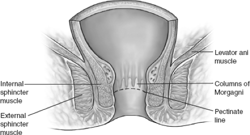
· Parts of the anal canal (AnatomyQA)

Related terms
References and sources
- Further reading: Pectinate line, Wikipedia.
- Further reading: Anal canal, Wikipedia.
- Federative International Programme for Anatomical Terminology (FIPAT). Terminologia Anatomica, 2nd ed. (TA2), 2019.