Anterior semicircular canal (Latin: canalis semicircularis anterior)
Also called Superior semicircular canal
· Of all parts of the labyrinth it lies closest to the bone surface, and it runs almost sagittally, at right angles to the long axis of the petrous part.
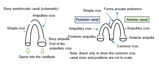
- Anterior (bony) semicircular canal : (= superior semicircular canal) the uppermost of the three. It runs almost sagittally and raises the arcuate eminence on the upper surface of the petrous part of the temporal bone. Its simple crus joins that of the posterior canal to form the (bony) common crus. End of the ampullary crus = anterior bony ampulla.length: about 15–20 mm
- Lateral (bony) semicircular canal : the shortest of the three and of middle width; it runs almost horizontally, backwards and laterally. Its bulge forms the prominence of the lateral semicircular canal on the medial wall of the tympanic cavity. End of the ampullary crus = lateral bony ampulla.length: about 12–15 mm
- Posterior (bony) semicircular canal : the lowest and longest of the three; it lies almost parallel to the long axis of the petrous part. Its simple crus joins that of the anterior canal to form the (bony) common crus. End of the ampullary crus = posterior bony ampulla.length: about 18–22 mm
- Anterior canal
- Simple crus : joins the simple crus of the posterior canal; as the (bony) common crus it opens into the medial wall of the back part of the vestibule.
- Ampullary crus → anterior bony ampulla : opens into the upper part of the vestibule, near the elliptical recess.
- Lateral canal
- Ampullary crus → lateral bony ampulla : opens into the vestibule just below the anterior ampulla, lateral to the elliptical recess.
- Simple crus : opens on its own into the back of the vestibule, between the common crus and the posterior ampulla.
- Posterior canal
- Simple crus : joins the simple crus of the anterior canal to form the common crus.
- Ampullary crus → posterior bony ampulla : opens into the lower back part of the vestibule.
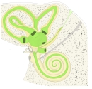 Bony and membranous labyrinth
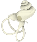 Bony labyrinth
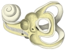 Bony labyrinth (opened)
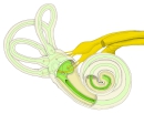 Labyrinth and nerves
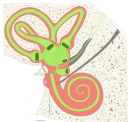 Perilymphatic space and membranous labyrinth
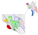 Right temporal bone from above
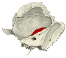 Arcuate eminence (right temporal bone, medial view)
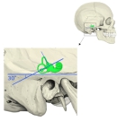 Position of the canals in the skull
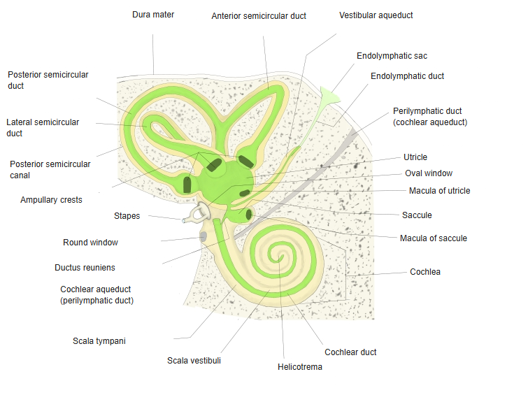
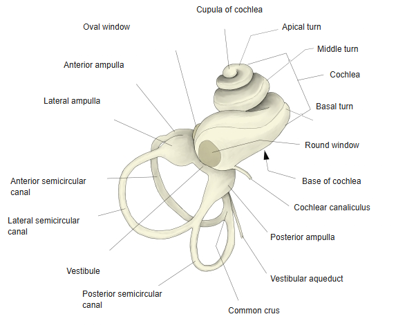
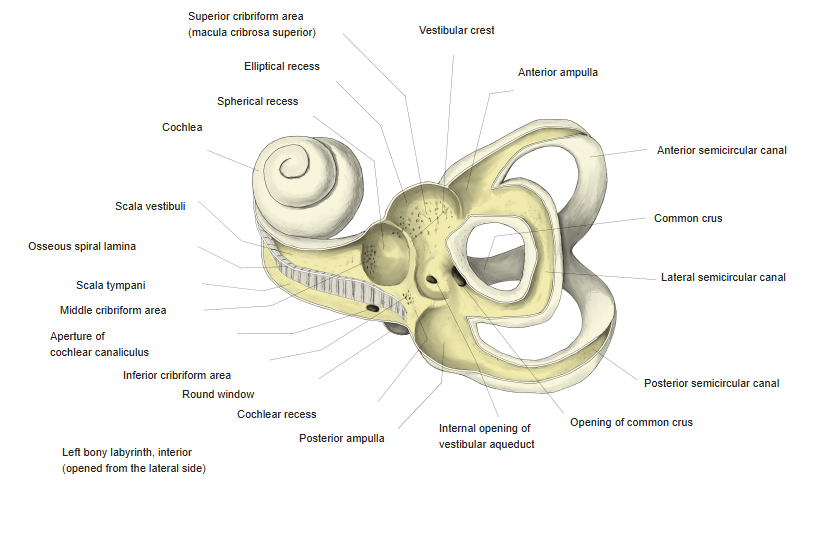
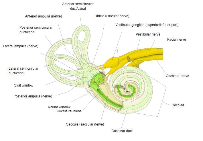
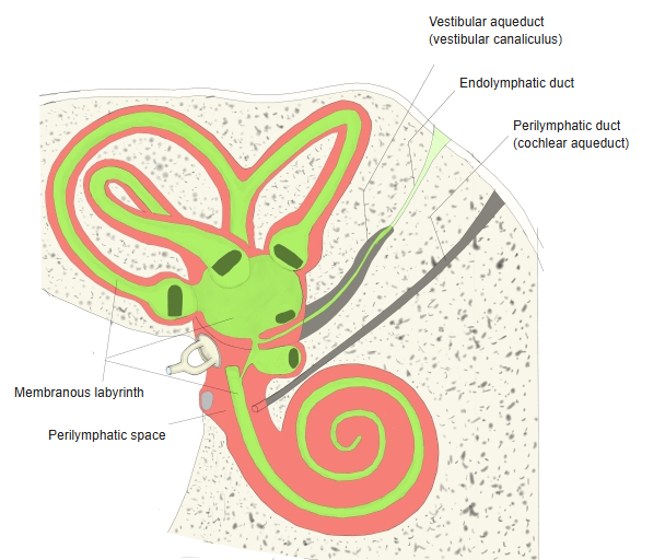
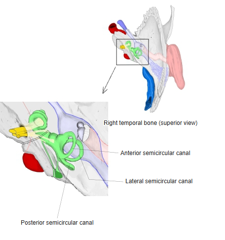
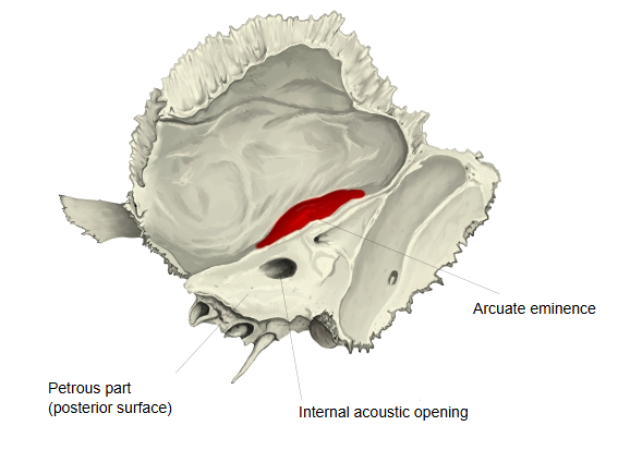
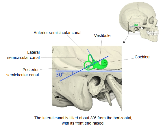
Because it lies closest to the bone surface and runs almost sagittally, its arch reaches up to just below the arcuate eminence on the upper surface of the petrous part. Its front (lateral) end begins with the ampullary crus at the anterior bony ampulla, which opens into the upper part of the vestibule; its back (medial) end forms the bony common crus together with the posterior canal.
· It contains the anterior semicircular duct and senses nodding movements of the head (rotation in the sagittal plane). The sensory crest in its ampulla is supplied by the anterior ampullary nerve. The other two canals are the posterior and the lateral semicircular canal.
· Thinning or absence of the bone over its arch (superior canal dehiscence) can cause vertigo set off by sound or pressure.
Reference websites
[Wikipedia]
· Semicircular canals, Wikipedia.
· Superior canal dehiscence syndrome, Wikipedia.
[Websites with illustrations]
· The inner ear: bony labyrinth and membranous labyrinth (TeachMeAnatomy)
· Internal ear anatomy: cochlea, vestibule and canals (Kenhub)
· Right osseous labyrinth, lateral view (Gray's Anatomy) (Wikimedia Commons)

Related terms
- Anterior bony ampulla
- Semicircular canals
- Semicircular ducts
- Posterior semicircular canal
- Lateral semicircular canal
- Bony labyrinth
- Vestibule
- Petrous part
References and sources
- Further reading: Semicircular canals, Wikipedia.
- Further reading: Superior canal dehiscence syndrome, Wikipedia.
- Federative International Programme for Anatomical Terminology (FIPAT). Terminologia Anatomica, 2nd ed. (TA2), 2019.