Latin: cavitas tympani
Overview
- The main space of the middle ear: a narrow air-filled cleft in the petrous part of the temporal bone, between the tympanic membrane and the internal ear.
- About 15 mm high and long, but only 2–6 mm wide.
- Contains the auditory ossicles, the tendons of the tensor tympani and stapedius, and the chorda tympani.
- Opens in front into the auditory tube and behind, through the aditus, into the mastoid antrum.
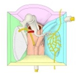
Walls of the tympanic cavity (model) 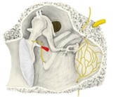
Tympanic cavity, medial wall
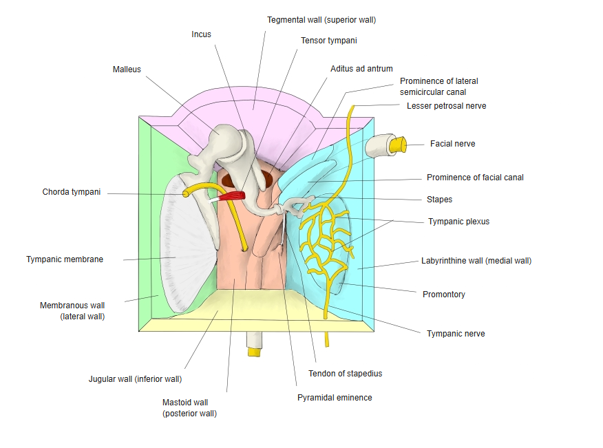
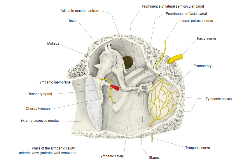
Walls
| Wall | Main features |
|---|---|
| Tegmental wall (roof) | Thin plate of bone (tegmen tympani) separating the cavity from the middle cranial fossa |
| Jugular wall (floor) | Thin bone over the bulb of the internal jugular vein; the tympanic nerve enters here |
| Membranous wall (lateral) | The tympanic membrane and, above it, the bony wall of the epitympanic recess |
| Labyrinthine wall (medial) | Promontory (bulge of the basal turn of the cochlea) with the tympanic plexus; oval window (closed by the base of the stapes) and round window; prominences of the facial canal and lateral semicircular canal |
| Mastoid wall (posterior) | Aditus to the mastoid antrum; pyramidal eminence containing the stapedius |
| Carotid wall (anterior) | Thin bone over the internal carotid artery; opening of the auditory tube and the canal for the tensor tympani |
Levels
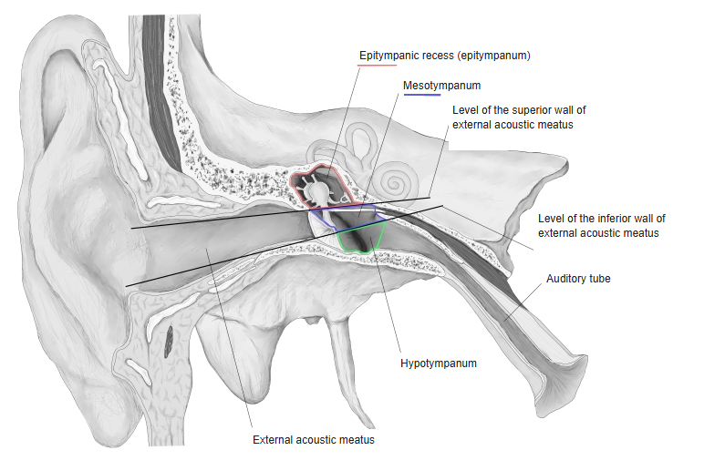
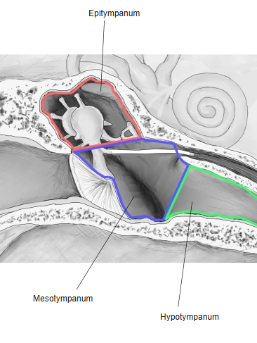
- The cavity is often divided by the level of the external acoustic meatus into three parts.
- Epitympanum (epitympanic recess): above the tympanic membrane; holds the head of the malleus and the body of the incus.
- Mesotympanum: directly medial to the tympanic membrane.
- Hypotympanum: a shallow space below the level of the membrane.
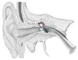
Levels of the tympanic cavity 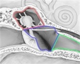
Epi-, meso- and hypotympanum (enlarged)
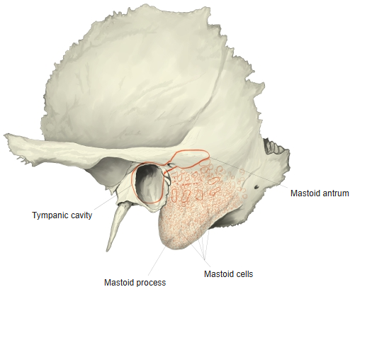
Mastoid antrum and mastoid cells
- The mastoid antrum is an air space behind the epitympanic recess, connected to it by the aditus.
- It opens into the mastoid cells, many small air spaces in the mastoid process; they develop mainly after birth.
- Infection of the middle ear can spread to them (mastoiditis).
Mastoid antrum and mastoid cells (schematic)
Blood supply and nerves
- Arteries: anterior tympanic (maxillary artery), stylomastoid (posterior auricular artery), superior tympanic (middle meningeal artery), inferior tympanic (ascending pharyngeal artery) and caroticotympanic arteries.
- Veins drain to the pterygoid plexus and the superior petrosal sinus.
- The mucous membrane is supplied by the tympanic plexus on the promontory, formed mainly by the tympanic nerve (glossopharyngeal nerve).
References and sources
- Further reading: Tympanic cavity, Wikipedia.