Bone lamellae (Latin: lamellae osseae)
Also called the lamellae of bone
· In neighbouring lamellae the collagen fibres run in different directions, which makes the bone strong in many directions, like plywood.
· In the shaft of a long bone four kinds of lamellae are distinguished:
lamellae
- Outer circumferential lamellae : the outermost lamellae of compact bone, just under the periosteum.
- Haversian lamellae : concentric rings around a central (Haversian) canal; together they form an osteon.
- Interstitial lamellae : fill the spaces between the osteons.
- Inner circumferential lamellae : the innermost lamellae of compact bone, next to the medullary cavity.
Osteocytes lie in small spaces (lacunae) between the lamellae and are linked by fine canals (canaliculi).
In spongy bone the lamellae run more or less parallel to the surface of each trabecula.
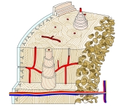 Lamellae of compact bone
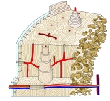 Structure of compact bone
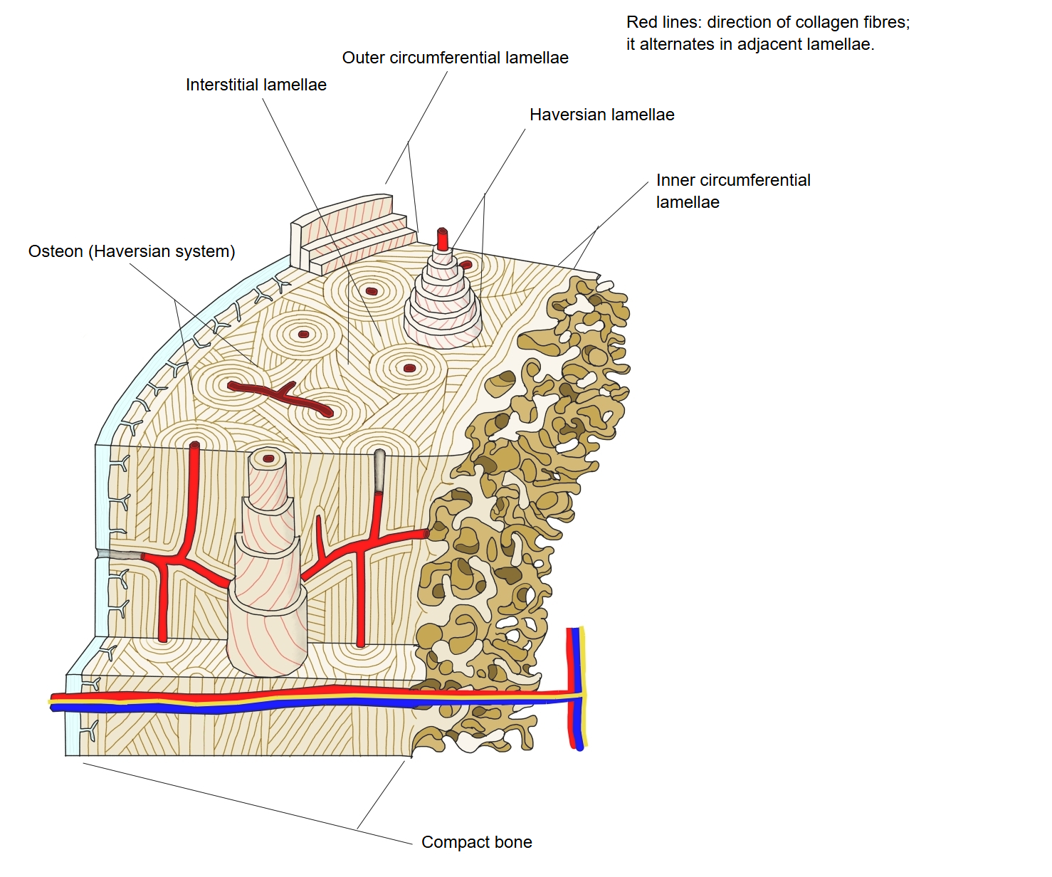
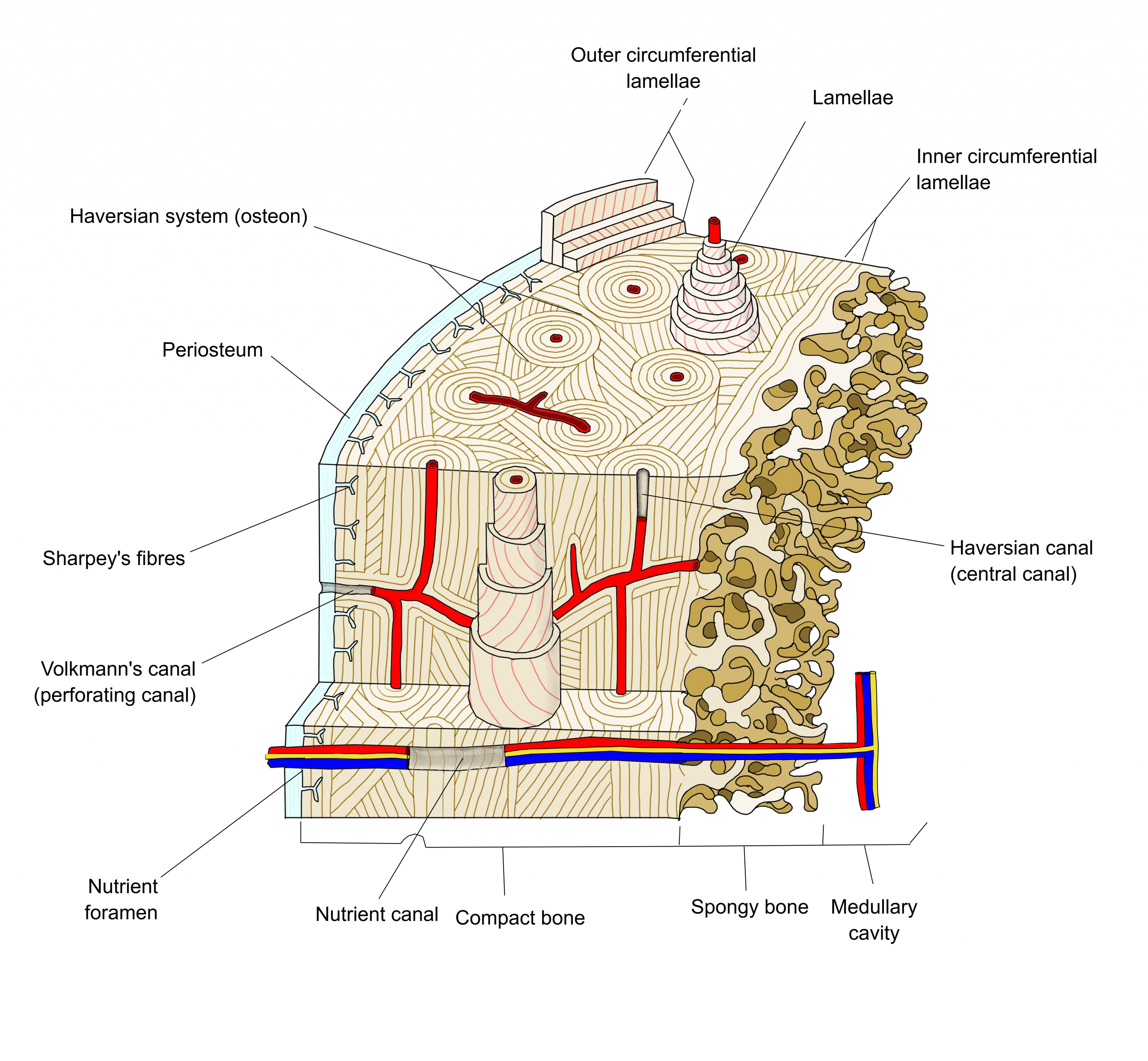
In mature (secondary) bone the lamellae are plates of collagen fibres embedded in the mineralized bone matrix, each only a few micrometres thick. Some lamellae lie parallel to one another; others are arranged in rings around a vascular canal, forming a Haversian system or osteon.
Reference websites
[Wikipedia]
· Osteon, Wikipedia.
· Bone, Wikipedia.
[Websites with illustrations]
· Cross-section of bone (Wikimedia Commons)

· Compact bone, histology (Wikimedia Commons)

· Bone structure (OpenStax Anatomy and Physiology 2e)
Related terms
- Bone trabeculae
- Osteon
- Interstitial lamellae
- Outer circumferential lamellae
- Inner circumferential lamellae
- Compact bone
- Bone matrix
- Perforating canal
- Spongy bone