Brush border
Also called the striated border
Overview
· The brush border is the edge formed on the luminal surface of epithelial cells by microvilli packed tightly side by side. In the light microscope the single microvilli cannot be told apart, so the edge looks like a band with fine vertical stripes; it is named for its likeness to the bristles of a brush.
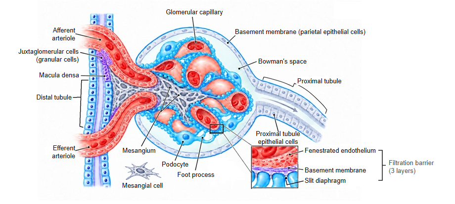
· In the kidney it is very well developed on the epithelium of the proximal tubule (the proximal convoluted tubule and the proximal straight tubule) and is the key feature for recognizing it. The distal tubule and the collecting duct have no brush border. Seen from the lumen, the edge shows delicate radial striations.
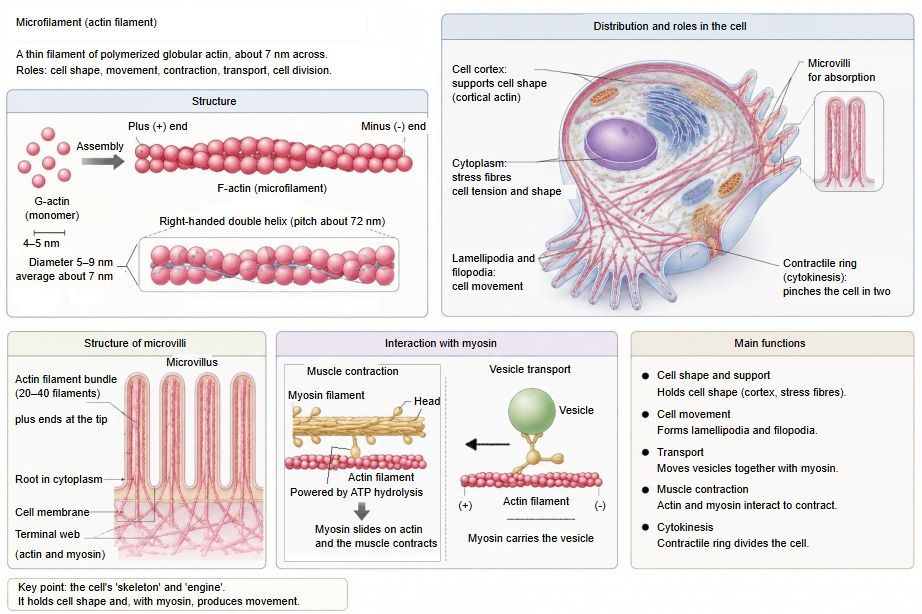
· Each microvillus is a slender projection about 0.1 µm across, with a core bundle of actin filaments (microfilaments). The microvilli greatly enlarge the cell surface and make the reabsorption of glucose, amino acids, sodium and other substances from the primary urine efficient.
· The surface of the absorptive cells of the small intestine (enterocytes) has the same structure, often called the striated border. The membrane of the intestinal microvilli carries enzymes for the last stage of digestion, such as the disaccharidases.
· Because of its carbohydrate surface coat it stains strongly with PAS. When the proximal tubule is injured, for example by ischaemia, the brush border is lost.
Reference websites
[Wikipedia]
· Brush border, Wikipedia.
· Microvillus, Wikipedia.
[Websites with illustrations]
· Microvilli of the duodenum (electron micrograph) (Wikimedia Commons)

· Kidney, CD10 staining of the proximal tubule brush border (Wikimedia Commons)

· Histology of the renal tubules (kidneypathology.com)
· Kidney (virtual slide) (Histology Guide)
Related terms
- Microvilli
- Proximal tubule
- Actin filament
- Enterocyte
- Renal tubule
- Distal tubule
- Proximal convoluted tubule
References and sources
- Further reading: Brush border, Wikipedia.
- Further reading: Microvillus, Wikipedia.
- Federative International Programme for Anatomical Terminology (FIPAT). Terminologia Anatomica, 2nd ed. (TA2), 2019.