Microvilli (Latin: microvilli)
Also called Microvillus (singular) or the brush border or the striated border
- Overview
- Microvilli outside the small intestine
- Microvilli, villi and cilia compared
- Reference websites
- (Wikipedia, websites with illustrations)
- Where it is found
- Related terms
Overview
Each microvillus is about 0.1 µm wide, so it can only be seen properly with the electron microscope; with the light microscope a dense row of them appears as the brush border (striated border). Each has a core of about 20–30 parallel actin filaments anchored in the terminal web just below the cell surface; microvilli are not motile in the way cilia are. Their surface carries a sugar-rich coat (glycocalyx) and digestive enzymes such as disaccharidases and peptidases, which finish digestion at the cell surface. Together with the circular folds and the villi they raise the absorptive surface of the small intestine many times over.
· In the small intestine, cells other than the absorptive intestinal epithelial cells also carry microvilli (goblet cells, enteroendocrine cells), but
they have very few short microvilli, which add little to the absorptive surface.
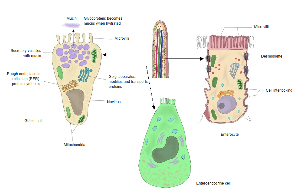
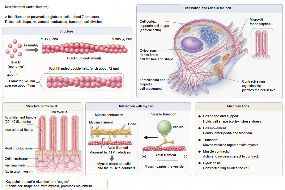
Microvilli outside the small intestine
| Organ or site | Role of the microvilli | Features | |
|---|---|---|---|
| 1 | Kidney (proximal tubule) | Reabsorption of water, salts, glucose and amino acids | A very well-developed, tall and dense brush border |
| 2 | Endometrium | Absorption and secretion | Their development changes with the menstrual cycle |
| 3 | Gallbladder | Absorption of water | Short microvilli enlarge the surface and help concentrate the bile |
| 4 | Part of the epithelium of the bronchi and other airways | Support of secretion | Distinct from the cilia of the same epithelium |
| 5 | Ducts of the pancreas (pancreatic duct) and salivary glands | Secretion | Present only in places |
| 6 | Large intestine | Absorption of water and salts | Short microvilli on the absorptive cells |
| 7 | Placenta | Exchange with the mother's blood | On the syncytiotrophoblast |
| 8 | Sensory cells | Detection of stimuli | The stereocilia of the hair cells of the inner ear and the tips of taste cells are specialized, very long or modified microvilli |
Microvilli, villi and cilia compared
| Structure | Size and shape | Function | Typical sites | |
|---|---|---|---|---|
| 1 | Microvilli | Fine projections of a single cell (seen with the electron microscope); actin core, not motile. Diameter 0.08–0.1 µm, length about 1–2 µm | Absorption, secretion | Small intestine, kidney |
| 2 | Villi | Large finger-like projections of the mucosa, made of many cells (seen with the naked eye or the light microscope). Diameter 0.1–0.3 mm, length 0.5–1.5 mm | Absorption | Small intestine only |
| 3 | Cilia | Motile projections of a single cell; microtubule core. Diameter about 0.2–0.25 µm, length about 5–10 µm | Moving mucus and other material along the surface | Airway epithelium, epithelium of the uterine tube |
Reference websites
[Wikipedia]
· Microvillus, Wikipedia.
· Brush border, Wikipedia.
[Websites with illustrations]
· Villi and microvilli of the small intestine (Wikimedia Commons)

· Microvilli of an intestinal cell (scanning electron micrograph) (Science Photo Library)
Where it is found
Related terms
References and sources
- Further reading: Microvillus, Wikipedia.
- Further reading: Brush border, Wikipedia.
- Federative International Programme for Anatomical Terminology (FIPAT). Terminologia Anatomica, 2nd ed. (TA2), 2019.