Collecting duct (Latin: ductus colligens)
Overview
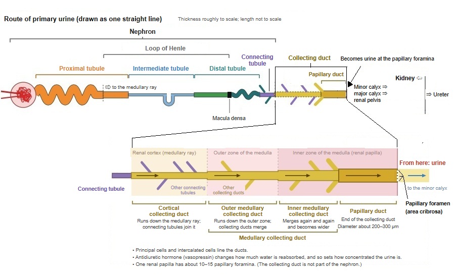
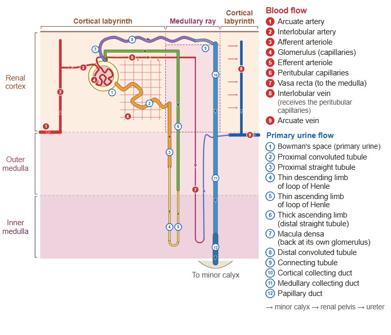
· The collecting duct receives the primary urine from the end of a nephron (the connecting tubule that follows the distal tubule) and carries it to the tip of the renal papilla.
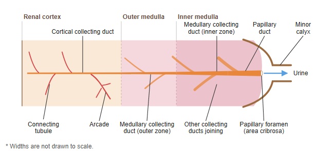
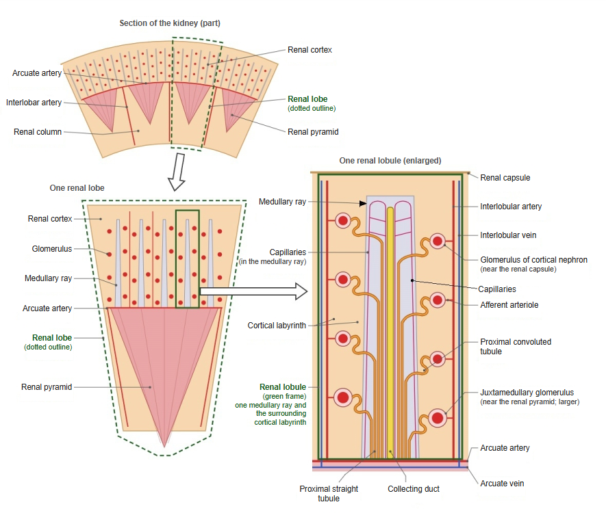
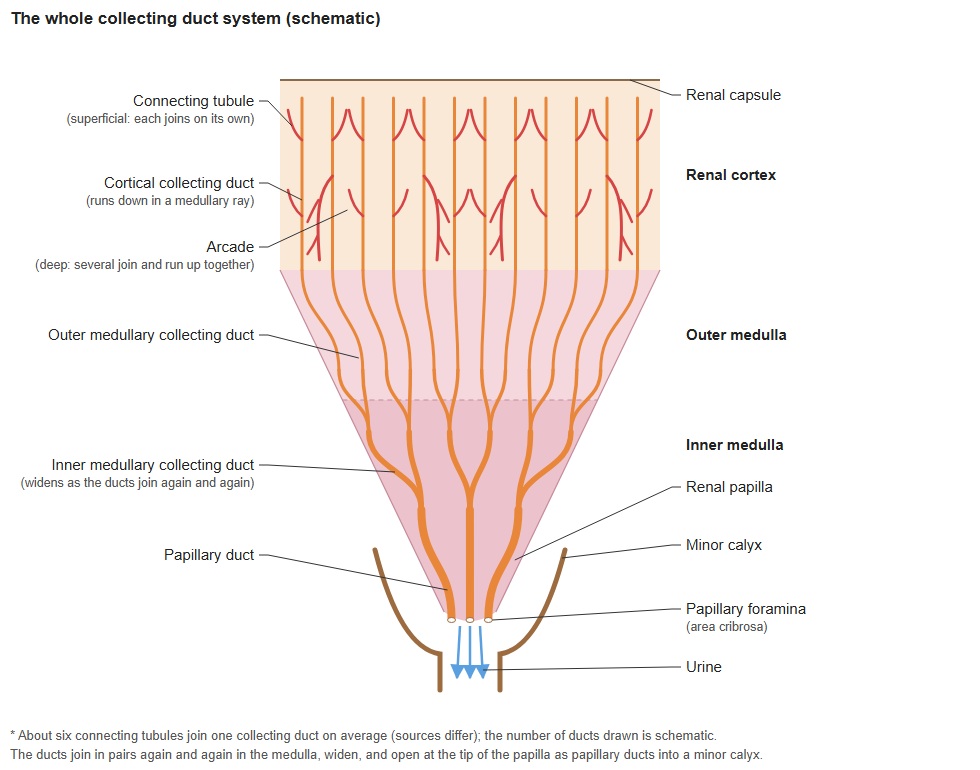
· It begins in a medullary ray of the renal cortex and runs down through the renal medulla (the renal pyramid) while many ducts join together.
· Its main parts are the cortical collecting duct and the medullary collecting duct. It ends as a wide papillary duct, which opens at the area cribrosa (the papillary foramina) on the tip of the papilla; there the fluid passes into a minor calyx as urine.
duct
- Cortical collecting duct : runs down in a medullary ray of the renal cortex.
- Medullary collecting duct : runs down through the renal medulla (outer and inner zones), joining other ducts and becoming wider.
- Papillary duct : releases the primary urine through the papillary foramina at the tip of the renal papilla into a minor calyx, where it becomes urine.
Note: the papillary duct is sometimes counted as part of the medullary collecting duct, as the wide final part of the inner medullary collecting duct.
of the
collecting duct
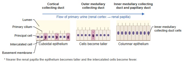
- Principal cells : reabsorb water (antidiuretic hormone) and Na⁺, and secrete K⁺ (aldosterone).
- Intercalated cells : secrete H⁺ or HCO₃⁻ and so regulate acid–base balance.
· On the way to the tip of the papilla several large branches unite to form each papillary duct, which opens on the tip of the papilla through a papillary foramen.
· The collecting duct has a different developmental origin from the nephron (it arises from the ureteric bud), so it is not counted as part of the nephron.
· Its wall is a cuboidal to columnar epithelium with principal cells and intercalated cells.
· Antidiuretic hormone (vasopressin) changes how much water is reabsorbed here and so sets the final concentration of the urine. Aldosterone regulates the reabsorption of sodium and the secretion of potassium, and the intercalated cells help to regulate acid–base balance.
- Renal cortex
- Renal corpuscle : glomerulus and Bowman's capsule.
- Proximal tubule : takes back water and nutrients from the primary urine.
- Distal tubule : adjusts electrolytes and water.
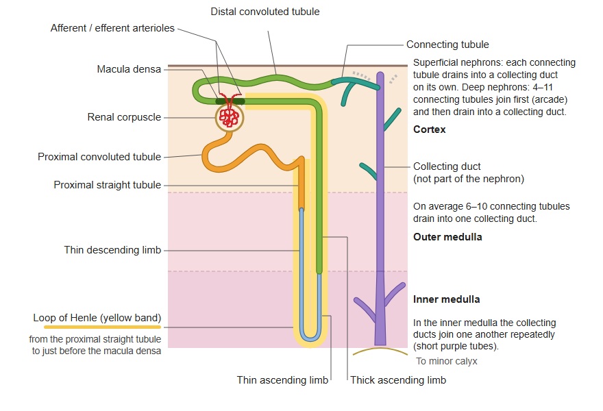
- Cortical collecting duct : collects the primary urine from several nephrons through their connecting tubules.
- Renal medulla
- Renal pyramid : the cone-shaped tissue that forms the medulla; it contains:
- Loop of Henle : part of the countercurrent system that concentrates the urine.
- Medullary collecting duct : carries the primary urine to the renal papilla.
- Papillary duct : opens at the tip of the papilla; its fluid passes into a minor calyx as urine.
- Renal pyramid : the cone-shaped tissue that forms the medulla; it contains:
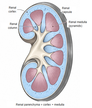
renal medulla (click to enlarge)
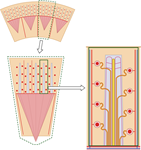 Renal lobe and renal lobule
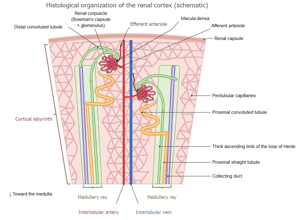
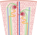 Tissue organization of the renal cortex
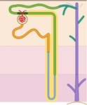 Nephron and collecting duct
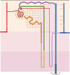 Nephron and blood vessels
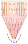 The whole collecting duct
Reference websites
[Wikipedia]
· Collecting duct system, Wikipedia.
[Websites with illustrations]
· The renal system (Nurse Key)
· Gray's Anatomy, figure 1128 (Wikimedia Commons)

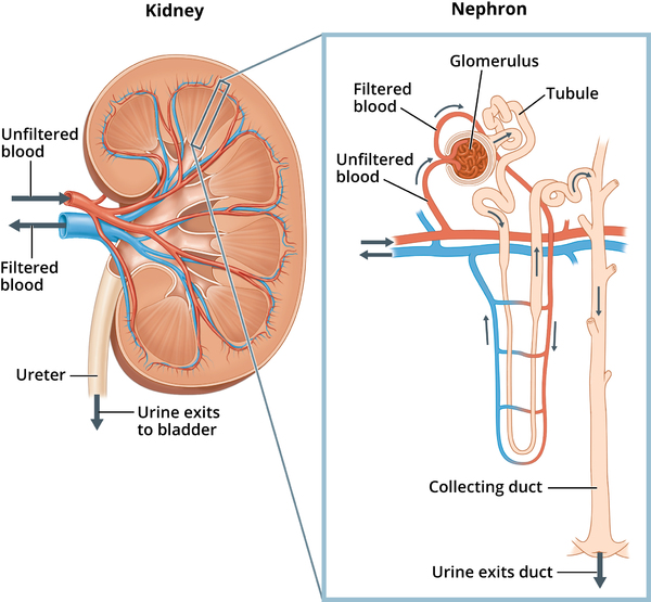
· Kidney and nephron (NIDDK)
Related terms
- Cortical collecting duct
- Medullary collecting duct
- Papillary duct
- Nephron
- Renal tubule
- Distal tubule
- Medullary rays
References and sources
- Further reading: Collecting duct system, Wikipedia.
- Federative International Programme for Anatomical Terminology (FIPAT). Terminologia Anatomica, 2nd ed. (TA2), 2019.