Central tendon of diaphragm (Latin: centrum tendineum)
· The central tendon of the diaphragm is the thin but strong sheet of tendon (aponeurosis) in the middle of the diaphragm. The muscle fibres of the surrounding parts (sternal, costal and lumbar) converge on it from all sides and insert into it.
Illustrations
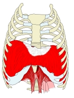 Diaphragm (anterior view)
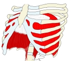 Diaphragm (anterolateral view)
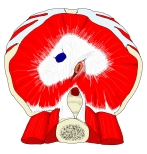 Diaphragm (inferior view)
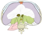 Diaphragm (inferior view, diagram)
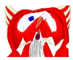 Diaphragm (inferior view)
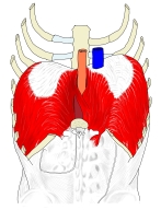 Diaphragm (posterior view)
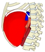 Diaphragm (right lateral view)
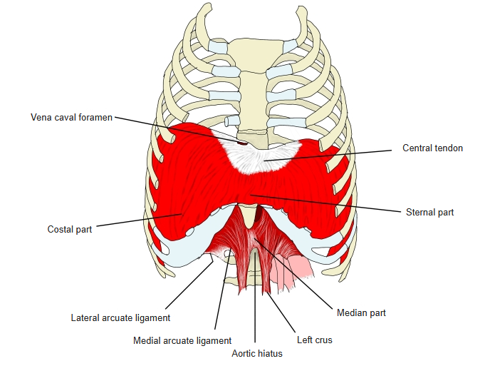
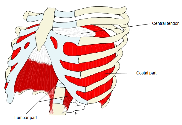
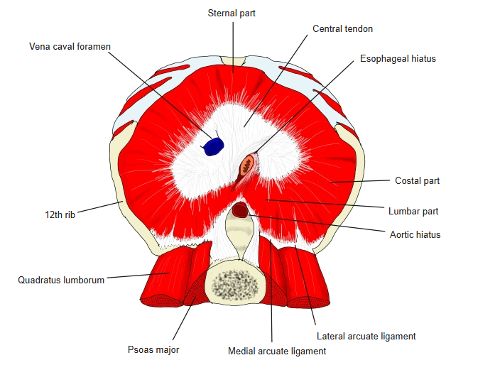
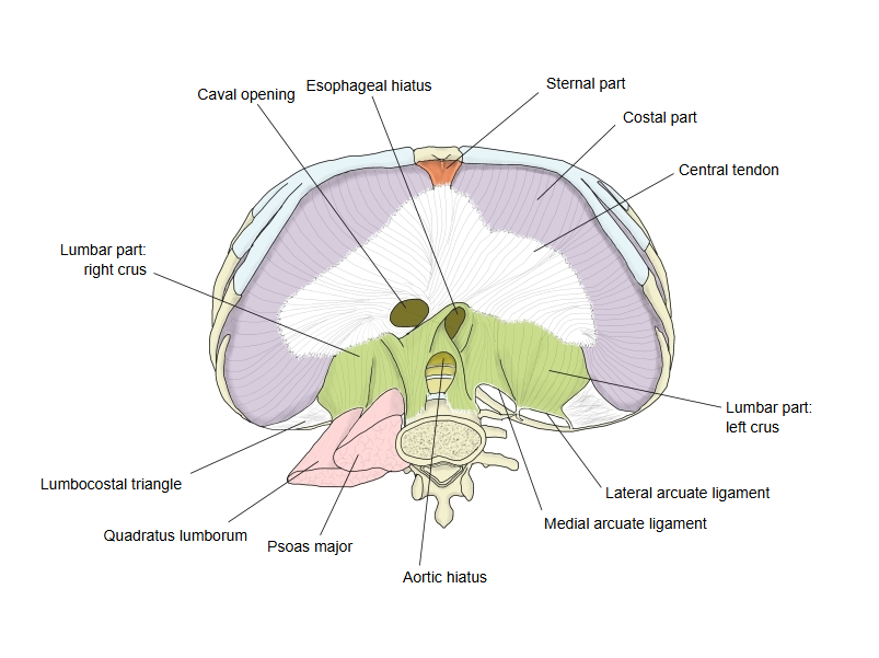
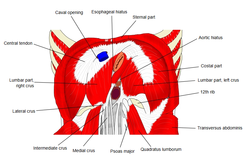
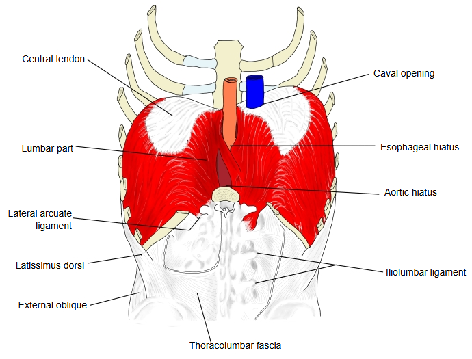
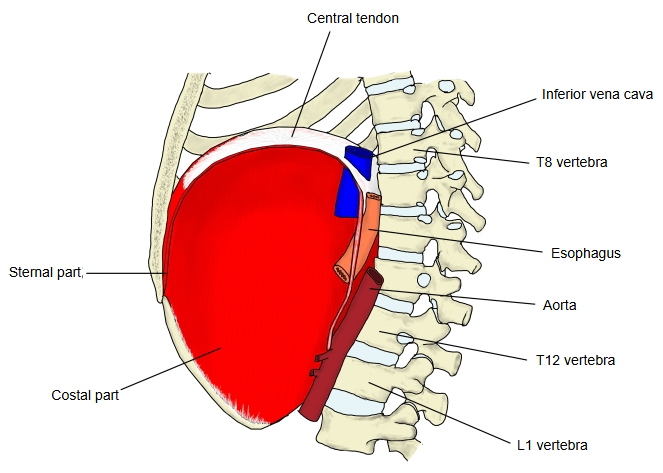
· As a whole it is shaped like a three-leaved clover, divided by shallow notches into right, middle and left leaflets. The right leaflet is the largest, the middle one (pointing forward toward the xiphoid process) comes next, and the left leaflet is the smallest.
· Its tendon fibres lie in several layers and cross one another at various angles to form bundles, which makes it very strong.
· Near the back edge of the right leaflet it is pierced by the caval opening (at about the level of the eighth thoracic vertebra), which carries the inferior vena cava and branches of the right phrenic nerve. The tendon bundles around the opening are firmly fixed to the wall of the vein, so the vein is always held open.
· Its upper surface is fused with the fibrous pericardium, and the heart rests on it. At the sides the upper surface is covered by the diaphragmatic pleura, and the lower surface by peritoneum.
· In breathing in, the diaphragm contracts and pulls the central tendon down, so the right and left domes of the diaphragm become a little flatter. The chest cavity gets larger, the pressure inside it falls and air flows into the lungs. Because the central tendon is tendon, it does not change shape and does not squeeze the inferior vena cava passing through the caval opening.
More details
· For the origin, insertion, action and nerve supply of the diaphragm as a whole, see Diaphragm.
Reference websites
[Wikipedia]
· Central tendon of diaphragm, Wikipedia.
· Thoracic diaphragm, Wikipedia.
[Websites with illustrations]
· Central tendon (IMAIOS e-Anatomy)
· The diaphragm (TeachMeAnatomy)
Related terms
- Diaphragm
- Inferior vena cava
- Fibrous pericardium
- Diaphragmatic pleura
- Phrenic nerve
- Parietal peritoneum
- Costodiaphragmatic recess
References and sources
- Further reading: Central tendon of diaphragm, Wikipedia.
- Further reading: Thoracic diaphragm, Wikipedia.
- Federative International Programme for Anatomical Terminology (FIPAT). Terminologia Anatomica, 2nd ed. (TA2), 2019.