Parietal peritoneum (Latin: peritoneum parietale)
Also called Parietal layer of peritoneum
What is the parietal peritoneum?
toneum
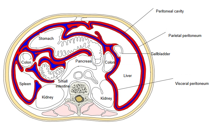
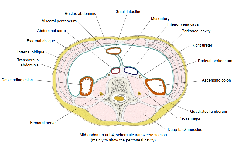
- Parietal peritoneum : the peritoneum covering the inner surface of the abdominal and pelvic cavities; also called the parietal layer.
- Visceral peritoneum : the peritoneum covering the surface of the organs in the abdominal and pelvic cavities; also called the visceral layer.
The two layers are continuous: they meet where the mesentery and the peritoneal ligaments leave the wall (see peritoneal cavity). Retroperitoneal organs such as the kidneys, the aorta and most of the duodenum lie behind the parietal peritoneum and are covered by it only on their front. The parietal peritoneum gets its blood and lymph from the body wall, and its nerves from the lower intercostal nerves, the subcostal, iliohypogastric and ilioinguinal nerves, and the phrenic nerve for the centre of the diaphragm; pain from it is therefore sharp and well localised. In peritonitis, irritation of the parietal layer causes local tenderness and reflex tightening (guarding) of the abdominal muscles.
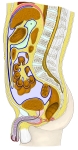 Abdomen and pelvis (median section)
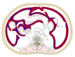 Abdomen (transverse section, schematic)
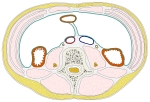 Mid-abdomen at L4 (transverse section)
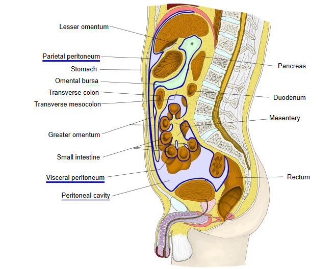
The medical dictionary of The Free Dictionary explains it simply as follows.
"the portion of the largest serous membrane in the body that lines the abdominal wall. Compare visceral peritoneum."
Useful sites
· Gray's Anatomy, figure 1040: lines along which the peritoneum leaves the abdominal wall to cover the organs (Wikimedia Commons)

· The Peritoneum (TeachMeAnatomy)
· Illustration of the peritoneum (image) (SlideShare)

Where it is found
Related terms
References and sources
- Further reading: Peritoneum, Wikipedia.
- Federative International Programme for Anatomical Terminology (FIPAT). Terminologia Anatomica, 2nd ed. (TA2), 2019.