Circular fibres of ciliary muscle (Latin: fibrae circulares musculi ciliaris)
Also called Müller's muscle, sphincter of the ciliary muscle
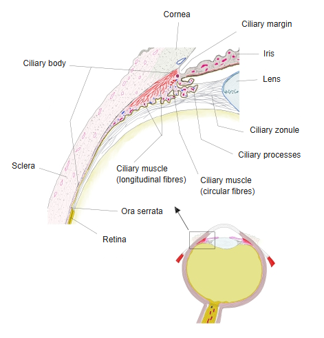
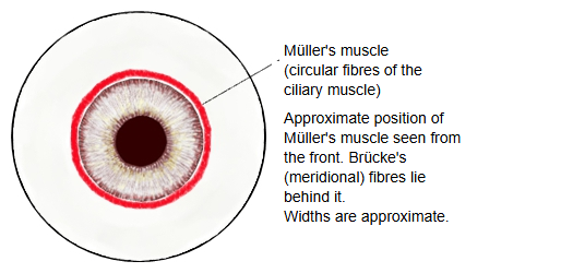
- Circular fibres : = Müller's muscle: ring-shaped fibres in the front inner part of the muscle; how well they are developed differs greatly between individuals.
- Meridional (longitudinal) fibres : = Brücke's muscle: run front to back from the boundary between the cornea and the sclera and reach the ciliary body.
- Radial (oblique) fibres : lie between the circular and meridional fibres and fan out from the boundary between the cornea and the sclera. Illustration of the three fibre groups (American Academy of Ophthalmology).
Contraction narrows the ring and moves the ciliary body toward the lens. The zonular fibres slacken and the lens becomes rounder for near vision (accommodation). The fibres are supplied by parasympathetic fibres from the oculomotor nerve through the ciliary ganglion and the short ciliary nerves. They tend to be well developed in long-sighted (hyperopic) eyes.
Note: the name Müller's muscle is also used for the superior tarsal muscle of the upper eyelid, a different smooth muscle. This page is about the ciliary muscle.
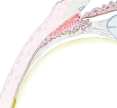 Section of the eyeball (front part enlarged)
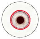 Müller's muscle (model)
Reference websites
[Wikipedia]
· Ciliary muscle, Wikipedia.
[Websites with illustrations]
· Longitudinal, radial and circular fibres of the ciliary muscle (American Academy of Ophthalmology)
Related terms
- Ciliary crown
- Ciliary ring
- Ciliary folds
- Ciliary processes
- Ciliary zonule
- Ciliary margin of iris
- Meridional fibres of ciliary muscle
- Radial fibers of ciliary muscle
- Ciliary body
- Lens
- Iris
- Ciliary ganglion
- Short ciliary nerves
References and sources
- Further reading: Ciliary muscle, Wikipedia.
- Federative International Programme for Anatomical Terminology (FIPAT). Terminologia Anatomica, 2nd ed. (TA2), 2019.