Ciliary ring (Latin: orbiculus ciliaris)
Also called the pars plana (flat part of the ciliary body)
· The ciliary crown in front is about 2-3 mm wide; the ciliary ring behind it is about 4 mm wide (sources differ on both widths).
- Ciliary crown (front)
- Ciliary processes : about 70 radial folds (sources give from about 60 to 90) rich in capillaries; they produce the aqueous humor.
- Ciliary folds : low folds between the ciliary processes.
- Ciliary ring (back) : a band about 4 mm wide that meets the ora serrata; many fine ridges run on its inner surface.
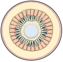 Ciliary body seen from inside
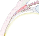 Ciliary body (section)
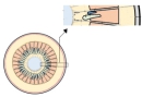 Ciliary crown and ciliary ring (enlarged)
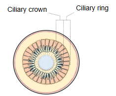
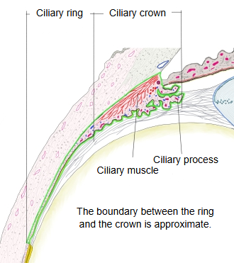
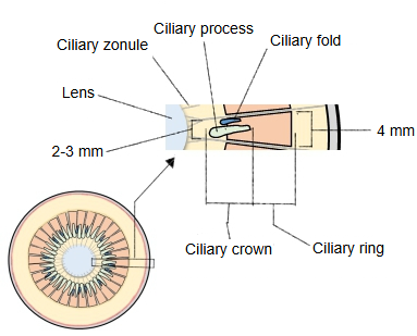
The back part of the ciliary body, about 4 mm wide, has shallow grooves and ridges running along the meridians; it is called the ciliary ring (pars plana). It is a little wider on the temporal side than on the nasal side, and its inner surface is covered by the two-layered epithelium of the ciliary part of the retina. The part in front of it, about 2-3 mm wide, is the ciliary crown (pars plicata): some 70 ciliary processes (sources differ on the number) project from it in a meridional pattern, and together with the ciliary folds between them they form a pleated ring like a crown. Fine fibres, the ciliary zonule, stretch between the ciliary body and the lens; many of them arise from the ciliary ring. All this is seen clearly when the front half of the eyeball is viewed from inside. Because the ciliary ring has few large vessels and lies in front of the light-sensitive retina, it is the usual entry site for instruments and injections into the vitreous body.
When the eyeball is cut at the equator and its front half is viewed from behind, the ciliary body can be seen all the way round the lens. Two zones are distinguished: (1) the ciliary ring, continuous with the ora serrata (its width is given differently in different descriptions; see above), and (2) the ciliary crown, made of about 70 radially arranged processes and the folds between them. Fine meridional ridges on the ring converge to form the ciliary processes (each about 2 mm long, 1 mm wide and 0.5 mm high).
Reference websites
[Wikipedia]
· Ciliary body, Wikipedia.
· Pars plana, Wikipedia.
[Websites with illustrations]
· Structure of the ciliary body (figure) (BrainKart)

· Ciliary body, processes and zonule (figure) (The Free Dictionary (medical))

Related terms
- Ciliary crown
- Ciliary folds
- Ciliary processes
- Ciliary zonule
- Ciliary margin of iris
- Circular fibres of ciliary muscle
- Ciliary body
- Ora serrata
- Retina
- Vitreous body
- Choroid
- Vascular layer of eyeball
References and sources
- Further reading: Ciliary body, Wikipedia.
- Further reading: Pars plana, Wikipedia.
- Federative International Programme for Anatomical Terminology (FIPAT). Terminologia Anatomica, 2nd ed. (TA2), 2019.