Fibular trochlea (Latin: trochlea fibularis)
Also called: peroneal trochlea; peroneal tubercle
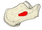 Right calcaneus (lateral)
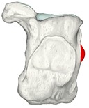 Right calcaneus (posterior)
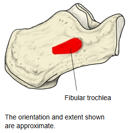
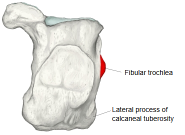
Anatomy textbooks describe it as follows.
On the lateral surface of the calcaneus runs a shallow, indistinct oblique groove, the groove for the tendon of fibularis longus. The small ridge just above it, long from front to back, is the fibular trochlea.
It lies on the lateral surface at about the junction of its front and middle thirds and runs obliquely downward and forward. Above it is a shallow groove for the tendon of fibularis brevis (the groove for the tendon of fibularis brevis); below it, the tendon of fibularis longus runs in its own groove.
Its size varies greatly from bone to bone. It is where the tendons of fibularis longus and brevis, which share a sheath behind the lateral malleolus, part to reach their separate attachments. The inferior fibular (peroneal) retinaculum attaches to it, and a large trochlea can irritate the tendons.
Reference websites
[Wikipedia]
· Calcaneus, Wikipedia.
· Fibularis longus, Wikipedia.
· Fibularis brevis, Wikipedia.
[Websites with illustrations]
· Calcaneus: anatomy and pathology (Kenhub)
· Bones of the foot: tarsals, metatarsals and phalanges (TeachMeAnatomy)
Where it is found
Related terms
- Groove for tendon of fibularis longus
- Body of calcaneus
- Lateral process of calcaneal tuberosity
- Calcaneal tuberosity
- Sustentaculum tali
- Calcaneus
References and sources
- Further reading: Calcaneus, Wikipedia.
- Further reading: Fibularis longus, Wikipedia.
- Further reading: Fibularis brevis, Wikipedia.
- Federative International Programme for Anatomical Terminology (FIPAT). Terminologia Anatomica, 2nd ed. (TA2), 2019.