Latin: calcaneus · also called the heel bone
Overview
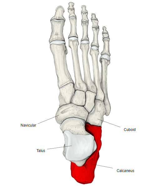
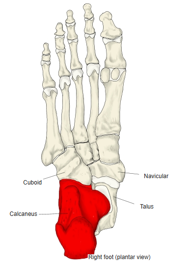
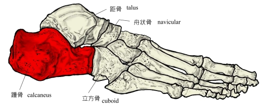
- One of the proximal tarsal bones of the foot and the largest of the tarsal bones: the bone of the heel.
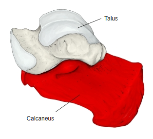
- It carries the talus on its upper surface.
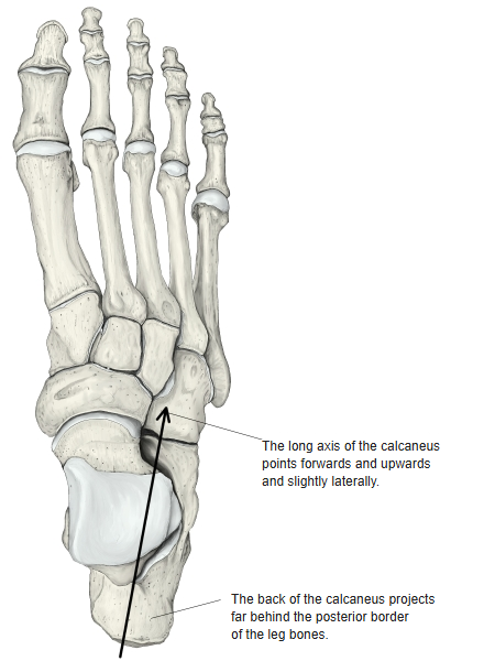
- An irregular, oblong block, long from front to back; its back part projects far behind the lower ends of the leg bones.
- Its long axis points forwards and upwards, and slightly laterally.
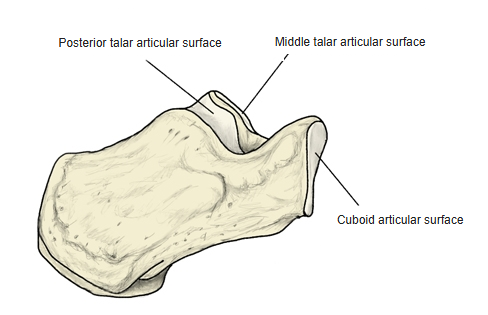
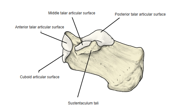
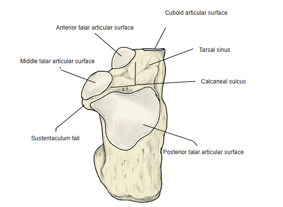
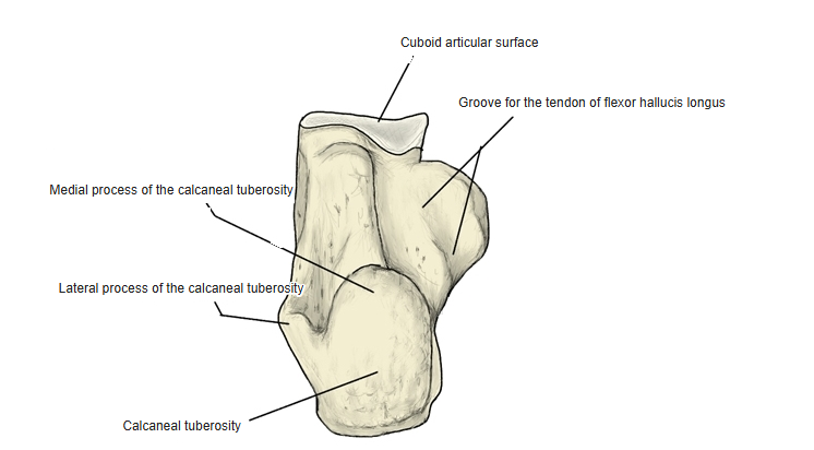
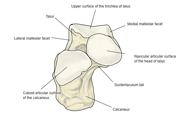
- Main features: the calcaneal tuberosity behind (with medial and lateral processes), the sustentaculum tali on the medial side, the fibular trochlea on the lateral side, the calcaneal sulcus (which forms the tarsal sinus with the talus), the talar articular surfaces (posterior, middle, anterior) above and the cuboid articular surface in front.
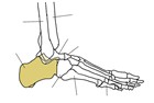
Right foot (lateral) 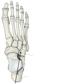
Long axis and heel
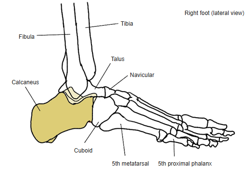
- Bones of the lower limb (one side)
- Pelvic girdle
- Hip bone ← ilium, ischium and pubis fused
- Free lower limb
- Thigh and knee
- Femur
- Patella
- Leg
- Tibia ← big-toe side
- Fibula ← little-toe side
- Foot
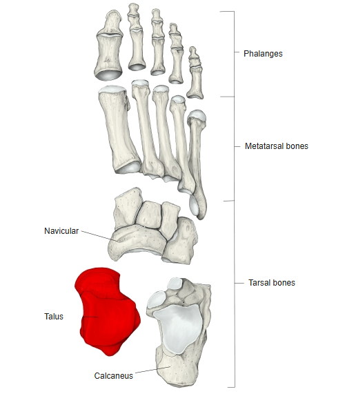
- Tarsal bones (7)
- Metatarsal bones (5)
- Phalanges (14)
- Thigh and knee
- Pelvic girdle

Illustrations
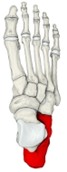
Right foot (dorsal) 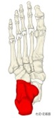
Right foot (plantar) 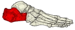
Right foot (lateral) 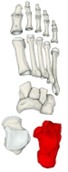
Bones of the foot (separated) 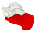
Talus and calcaneus (medial) 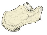
Right calcaneus (lateral) 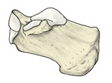
Right calcaneus (medial) 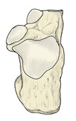
Right calcaneus (from above) 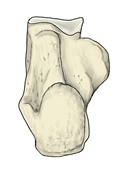
Right calcaneus (from below) 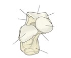
Talus and calcaneus (anterior) 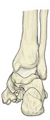
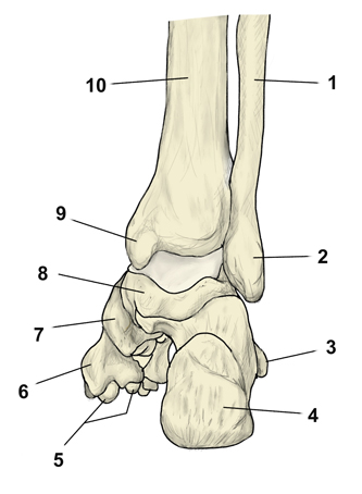
Right foot (posterior) 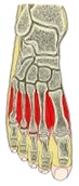
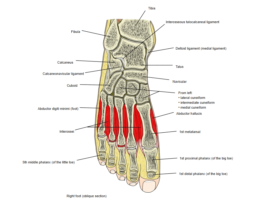
Right foot (section)
Articulations
| # | Bone | Joint |
|---|---|---|
| 1 | Talus | Subtalar (talocalcaneal) joint |
| 2 | Talus | Talocalcaneonavicular joint |
| 3 | Cuboid | Calcaneocuboid joint |
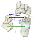
Joints of the calcaneus
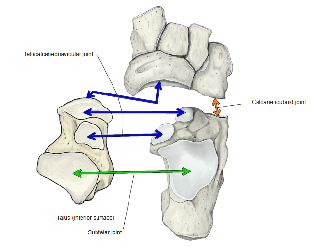
Subtalar joint: posterior talar articular surface ↔ posterior calcaneal articular surface of the talus. Talocalcaneonavicular joint: anterior and middle talar articular surfaces ↔ anterior and middle calcaneal articular surfaces of the talus (with the navicular). Calcaneocuboid joint: cuboid articular surface of the calcaneus ↔ calcaneal articular surface of the cuboid.
Ligaments
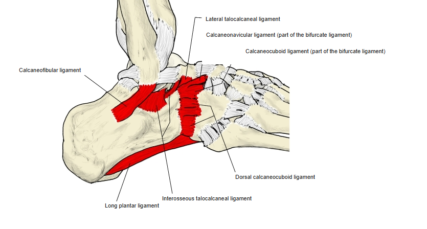
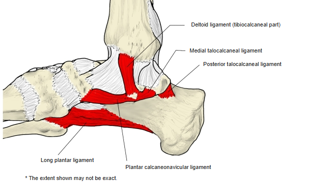
The following ligaments attach to the calcaneus. The list is intended to be comprehensive but may omit minor ligaments.
| # | Ligament | Connects |
|---|---|---|
| 1 | Lateral talocalcaneal ligament | Calcaneus – lateral process of talus (front part) |
| 2 | Medial talocalcaneal ligament | Sustentaculum tali – medial tubercle of talus |
| 3 | Interosseous talocalcaneal ligament | Calcaneal sulcus – sulcus tali |
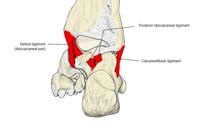
| 4 | Posterior talocalcaneal ligament | Calcaneus – medial and lateral tubercles of talus |
| 5 | Calcaneocuboid ligament (part of the bifurcate ligament) | Dorsal surface – dorsal surface of cuboid |
| 6 | Calcaneonavicular ligament (part of the bifurcate ligament) | Dorsal surface – navicular |
| 7 | Dorsal calcaneocuboid ligament | Dorsal surface – dorsal surface of cuboid |
| 8 | Deltoid ligament (tibiocalcaneal part) | Sustentaculum tali – medial malleolus of tibia |
| 9 | Calcaneofibular ligament | Lateral surface – lateral malleolus of fibula |
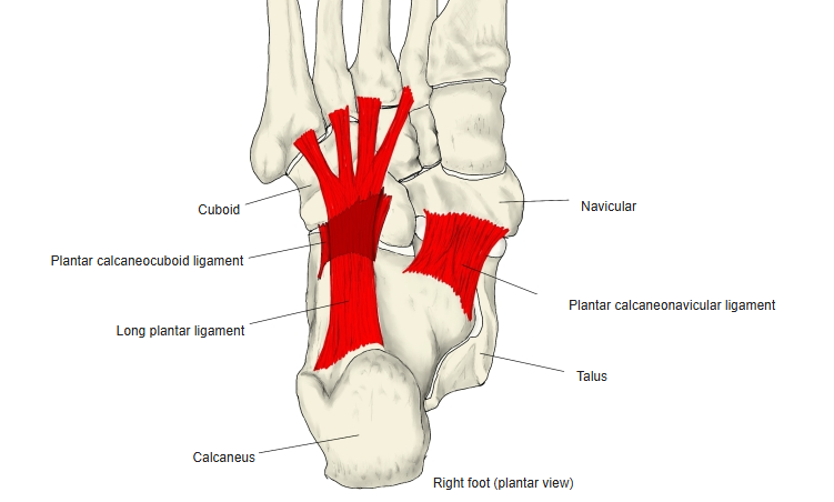
| 10 | Long plantar ligament | Plantar surface of the calcaneus (in front of the tuberosity) – tuberosity of cuboid and bases of the metatarsals |
| 11 | Plantar calcaneonavicular ligament | Sustentaculum tali – navicular (plantar surface, medial border, upper medial part) |
| 12 | Plantar calcaneocuboid ligament | Calcaneus (front of the plantar surface) – cuboid |
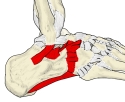
Right foot (lateral) 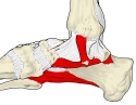
Right foot (medial) 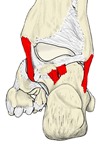
Right foot (posterior) 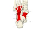
Right foot (plantar)
Muscle attachments
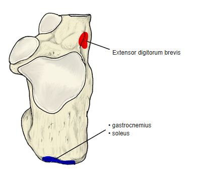
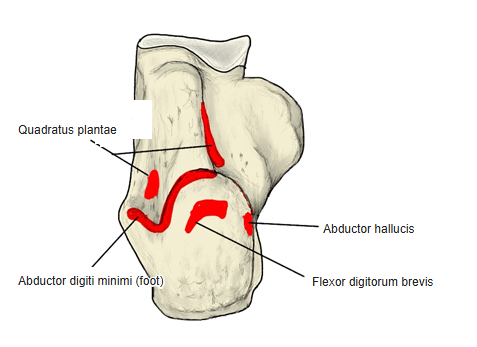
- Origin of: Abductor digiti minimi (foot), Quadratus plantae, Flexor digitorum brevis, Abductor hallucis, Extensor digitorum brevis
- Insertion of: Gastrocnemius, Soleus
Origins: abductor digiti minimi, flexor digitorum brevis and abductor hallucis — calcaneal tuberosity (plantar side); quadratus plantae — plantar surface; extensor digitorum brevis — forepart of the upper and lateral surfaces. Insertion: gastrocnemius and soleus — calcaneal tuberosity through the calcaneal (Achilles) tendon.
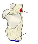
Muscle attachments (from above) 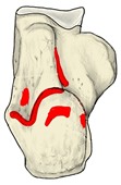
Muscle attachments (from below)
References and sources
- Further reading: Calcaneus, Wikipedia.