Frontal notch (Latin: incisura frontalis)
Also called the frontal foramen (when closed into a hole)
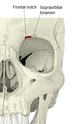
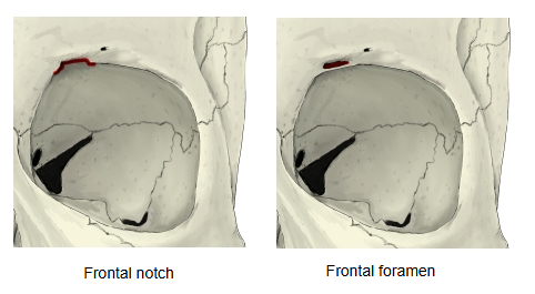
A notch on the medial part of the supraorbital margin of the frontal bone (medial to the supraorbital foramen or notch). The nerve and vessels listed below pass through it. When it is closed into a hole rather than a notch, it is called the frontal foramen (see the illustration below).
Artery: supratrochlear artery
Vein: supratrochlear vein
About the junction of the medial and middle thirds of the orbital margin there is a notch or hole, called in older texts the lateral frontal notch (foramen) and today the supraorbital notch (foramen). Medial to it is a groove, the medial frontal notch, which is sometimes closed into a medial frontal foramen: this is the frontal notch of current terminology. Laterally the orbital margin ends in a strong projection that joins the zygomatic bone, the zygomatic process.
Reference websites
[Wikipedia]
· Frontal bone, Wikipedia.
[Websites with illustrations]
· Frontal bone, outer surface (Gray's Anatomy, fig. 134) (Wikimedia Commons)

· Frontal bone: anatomy, borders and development (Kenhub)
Related terms
- Supra-orbital margin
- Supraorbital region
- Orbital part of frontal bone
- Nasal part of frontal bone
- Frontal bone
References and sources
- Further reading: Frontal bone, Wikipedia.
- Federative International Programme for Anatomical Terminology (FIPAT). Terminologia Anatomica, 2nd ed. (TA2), 2019.