Latin: os zygomaticum
Overview
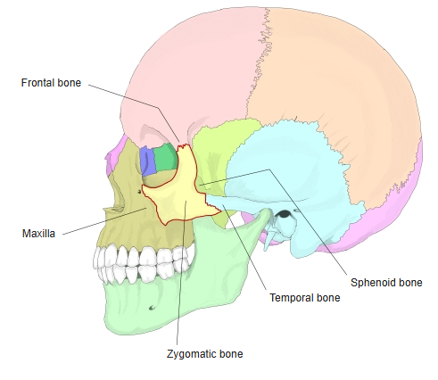
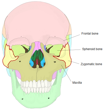
- The paired cheek bone, forming the prominence of the cheek, part of the lateral wall and floor of the orbit and the front of the zygomatic arch.
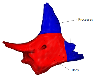
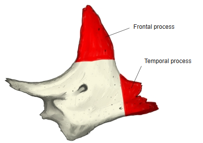
- It consists of a body and processes: the frontal process rises to meet the frontal bone, the temporal process runs back to meet the zygomatic process of the temporal bone, and a maxillary process joins the maxilla.
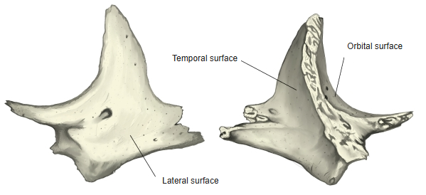
- It has three surfaces: lateral, temporal and orbital.
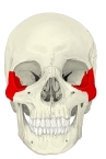
Skull (anterior) 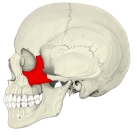
Skull (left lateral) 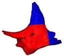
Body and processes
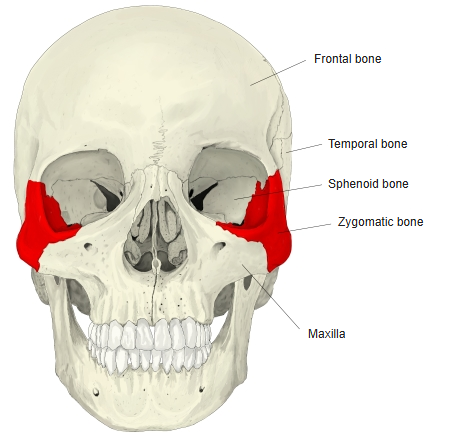
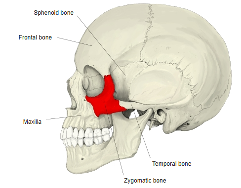
- Bones of the skull
- Neurocranium (cranial bones)
- Frontal bone
- Parietal bone (2)
- Occipital bone
- Temporal bone (2)
- Sphenoid bone
- Ethmoid bone
- Viscerocranium (facial bones)
- Nasal bone (2)
- Vomer
- Lacrimal bone (2)
- Inferior nasal concha (2)
- Maxilla (2)
- Zygomatic bone (2)
- Palatine bone (2)
- Mandible
- Hyoid bone
- Neurocranium (cranial bones)
Illustrations
Articulations
| # | Bone | Joint |
|---|---|---|
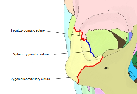
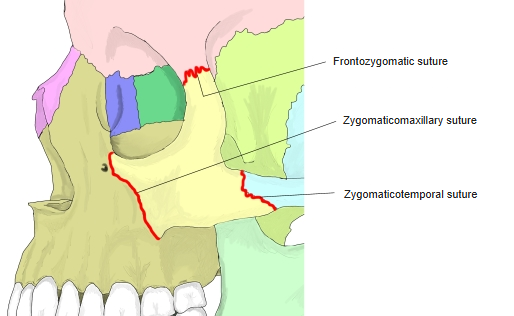
| 1 | Frontal bone | Frontozygomatic suture |
| 2 | Sphenoid bone (greater wing) | Sphenozygomatic suture |
| 3 | Maxilla | Zygomaticomaxillary suture |
| 4 | Temporal bone | Zygomaticotemporal suture |
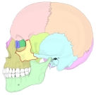
Neighbouring bones (lateral) 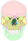
Neighbouring bones (anterior) 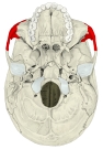
External cranial base 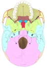
External cranial base (coloured)
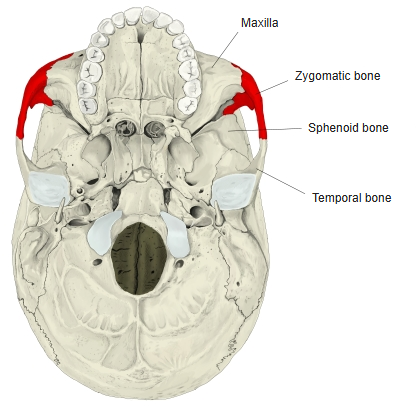
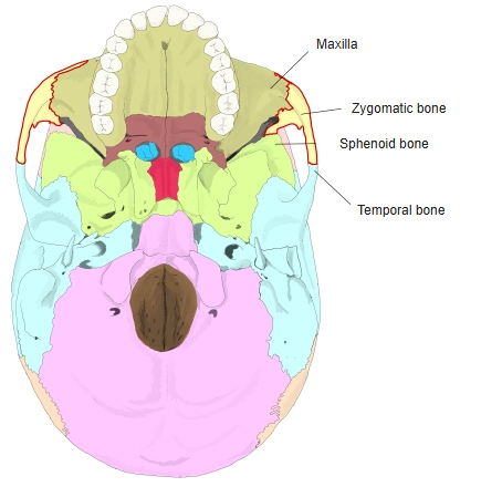
Muscle attachments
- Origin of: Zygomaticus major, Zygomaticus minor, Masseter (superficial part)
- Insertion of: none
Masseter arises from the lower border of the zygomatic arch; temporal fascia attaches to the upper border of the frontal and temporal processes. Some sources also give part of levator labii superioris as arising here.
Muscle attachments
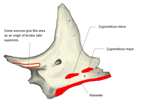
Surfaces and processes
- Lateral surface – convex, forming the cheek; the small zygomaticofacial foramen opens on it.
- Temporal surface – faces backwards and medially into the temporal and infratemporal fossae; the zygomaticotemporal foramen opens on it.
- Orbital surface – smooth and concave, forming part of the lateral wall and floor of the orbit; the zygomatico-orbital foramen opens on it.
- Frontal process – thick and serrated above, where it meets the zygomatic process of the frontal bone; the small marginal tubercle lies on its posterior border.
- Temporal process – joins the zygomatic process of the temporal bone to complete the zygomatic arch.
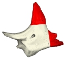
Frontal and temporal processes 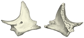
Surfaces
Sutures
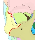
- Frontozygomatic suture – with the frontal bone.
- Sphenozygomatic suture – with the greater wing of the sphenoid, inside the orbit and in the temporal fossa.
- Zygomaticomaxillary suture – with the maxilla.
- Zygomaticotemporal suture (temporozygomatic suture) – with the temporal bone in the zygomatic arch.
Sutures (lateral) 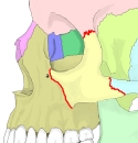
Sutures (anterolateral)
References and sources
- Further reading: Zygomatic bone, Wikipedia.