Guyon's canal
Also called the ulnar canal, ulnar tunnel
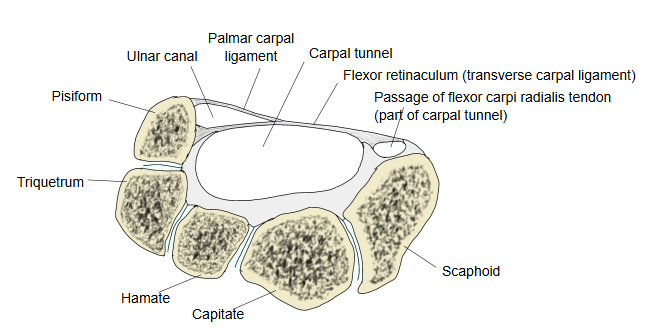
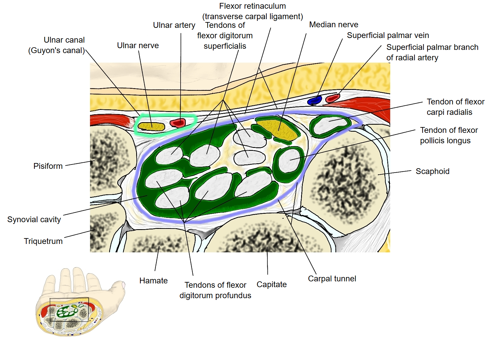
= the ulnar canal (ulnar tunnel): a short passage, about 4 cm long, on the little-finger side of the front of the wrist, through which the ulnar nerve and the ulnar artery enter the hand. Its roof is the palmar carpal ligament, a thickening of the deep fascia of the forearm (some sources include the palmaris brevis); its floor is the flexor retinaculum and the hypothenar muscles; its medial wall is the pisiform and the pisohamate ligament, and its lateral wall the hook of hamate. It lies superficial to and on the ulnar side of the carpal tunnel, which carries the median nerve and the long flexor tendons. Inside it the ulnar nerve divides into its superficial and deep branches; pressure on the nerve here (ulnar tunnel syndrome) can cause weakness of the small hand muscles and numbness of the little finger.
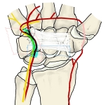 Ulnar canal at the wrist
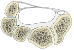 Ulnar canal and carpal tunnel
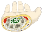 Cross-section of the wrist
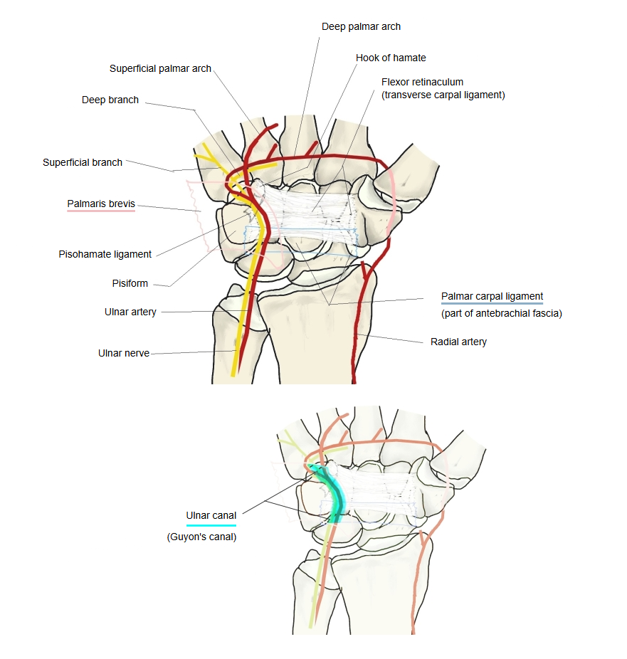
Related terms
- Carpal tunnel
- Flexor retinaculum (hand)
- Hook of hamate
- Pisiform
- Hypothenar muscles
- Ulnar nerve
- Ulnar artery
References and sources
- Further reading: Ulnar canal, Wikipedia.
- Federative International Programme for Anatomical Terminology (FIPAT). Terminologia Anatomica, 2nd ed. (TA2), 2019.