Head of femur (Latin: caput femoris)
Also called the femoral head
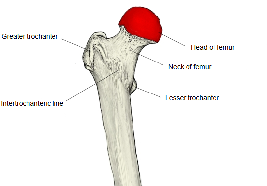
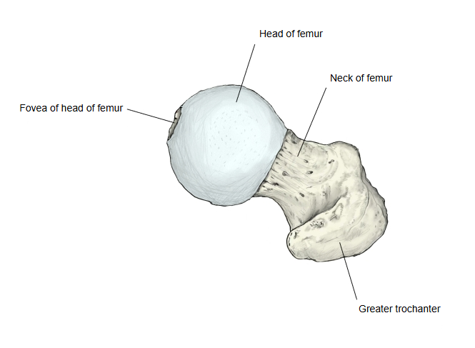
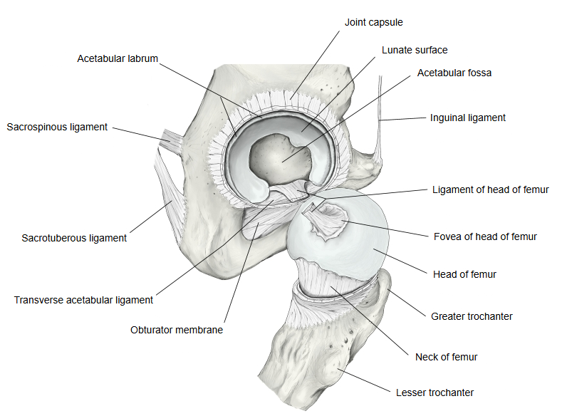
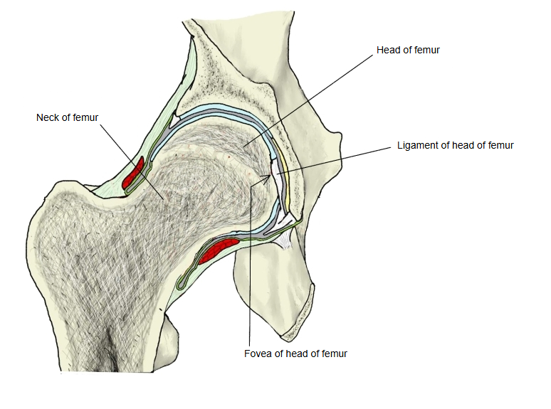
The nearly spherical, cartilage-covered part of the upper end of the femur, at the end of the neck of femur, which projects upwards and medially. It forms a little more than half a sphere and faces upwards, medially and slightly forwards. Covered with articular cartilage, it meets the lunate surface of the acetabulum of the hip bone to form the hip joint. A little below and behind its centre is the fovea of head of femur, a small pit without cartilage, where the ligament of head of femur is attached. Most of the blood supply of the adult head reaches it along the neck, so fractures of the neck can lead to avascular necrosis of the head.
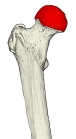 Right femur (upper end, anterior)
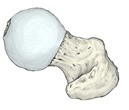 Right femur (upper end, from above)
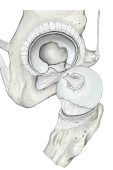 Right hip joint (opened)
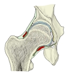 Right hip joint (coronal section)
Reference websites
[Wikipedia]
· Femoral head, Wikipedia.
[Websites with illustrations]
· Head of femur with its articular cartilage (photo) (Wikimedia Commons)

· The Femur (TeachMeAnatomy)
Where it is found
Related terms
- Fovea of head of femur
- Neck of femur
- Proximal epiphysis of femur
- Greater trochanter
- Hip joint
- Hip bone
References and sources
- Further reading: Femoral head, Wikipedia.
- Further reading: Hip, Wikipedia.
- Federative International Programme for Anatomical Terminology (FIPAT). Terminologia Anatomica, 2nd ed. (TA2), 2019.