Latin: femur · also called the thigh bone
Overview
- The only bone of the thigh and the longest tubular bone of the body: about 40 cm, roughly a quarter of body height.
- It is slightly convex anteriorly.
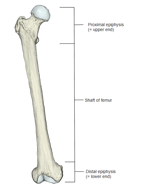
- It has a shaft and two ends: the proximal (upper) end and the distal (lower) end.
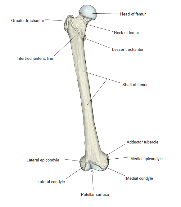
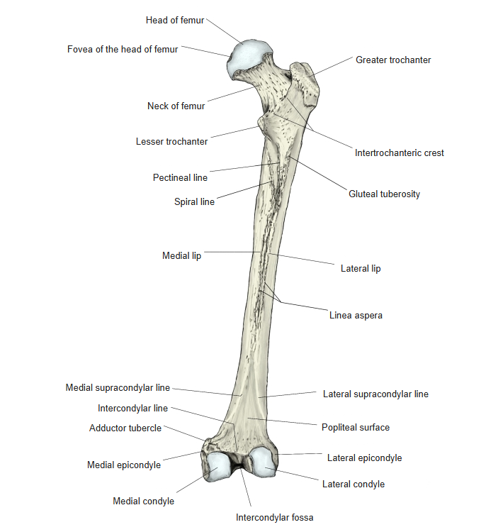
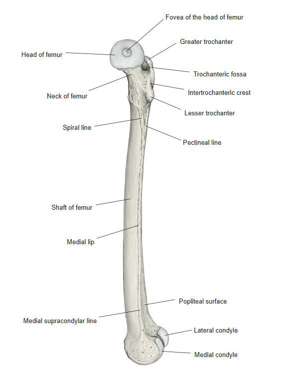
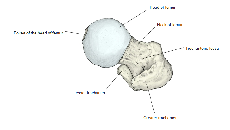
- Upper end: the head of the femur (with the fovea), the neck, the greater and lesser trochanters, the intertrochanteric line in front and the intertrochanteric crest behind.
- Shaft: the linea aspera on the back (medial and lateral lips), the gluteal tuberosity and the pectineal line.
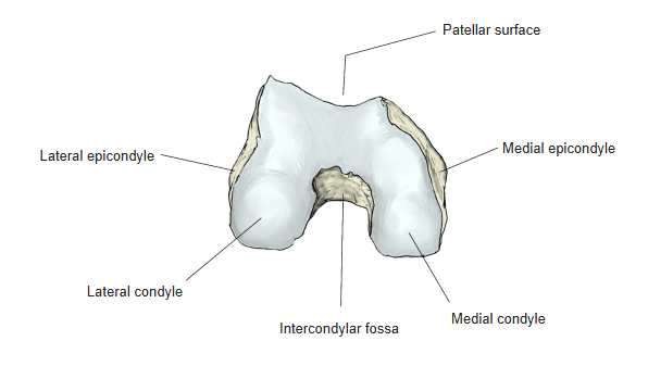
- Lower end: the medial and lateral condyles with the intercondylar fossa between them, the epicondyles, the adductor tubercle and the patellar surface in front.
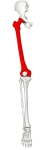
Right femur 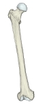
Parts of the femur
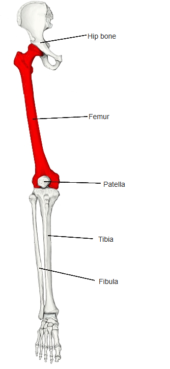
- Bones of the lower limb (one side)
- Pelvic girdle
- Hip bone ← ilium, ischium and pubis fused
- Free lower limb
- Thigh and knee
- Femur
- Patella
- Leg
- Tibia ← big-toe side
- Fibula ← little-toe side
- Foot
- Tarsal bones (7)
- Metatarsal bones (5)
- Phalanges (14)
- Thigh and knee
- Pelvic girdle
Illustrations
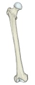
Right femur (anterior) 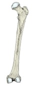
Right femur (posterior) 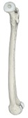
Right femur (medial) 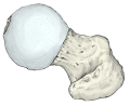
Upper end, seen from above 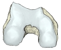
Lower end, seen from below
Articulations
| # | Bone | Joint |
|---|---|---|
| 1 | Hip bone | Hip joint |
| 2 | Tibia | Knee joint |
| 3 | Patella |
Joints of the femur
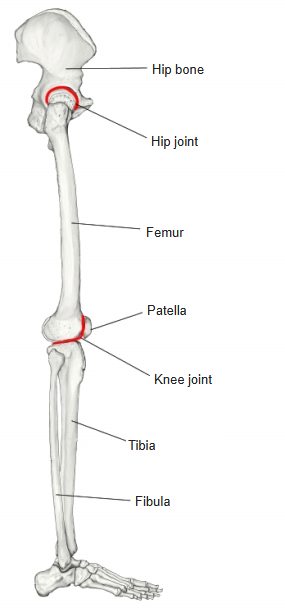
Hip joint: head of the femur ↔ acetabulum of the hip bone. Knee joint: medial and lateral condyles of the femur ↔ medial and lateral condyles of the tibia, and patellar surface of the femur ↔ posterior surface of the patella. Some sources describe the femur–patella part separately as the patellofemoral joint.
Ligaments
The following ligaments attach to the femur. The list is intended to be comprehensive but may omit minor ligaments.
| # | Ligament | Connects |
|---|---|---|
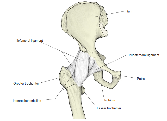
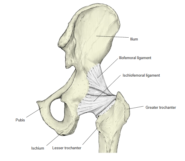
| 1 | Iliofemoral ligament | Intertrochanteric line – anterior inferior iliac spine and upper rim of the acetabulum |
| 2 | Pubofemoral ligament | Lesser trochanter – iliopubic eminence and superior pubic ramus |
| 3 | Ischiofemoral ligament | Neck of femur and trochanteric fossa – rim of the acetabulum (ischium) |
| 4 | Ligament of the head of femur | Fovea of the head of femur – acetabular fossa |
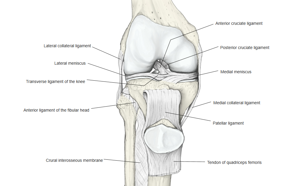
| 5 | Fibular (lateral) collateral ligament | Lateral epicondyle – head of fibula |
| 6 | Tibial (medial) collateral ligament | Medial epicondyle – medial condyle of tibia |
| 7 | Anterior cruciate ligament | Lateral condyle – anterior intercondylar area of tibia |
| 8 | Posterior cruciate ligament | Medial condyle – posterior intercondylar area of tibia |
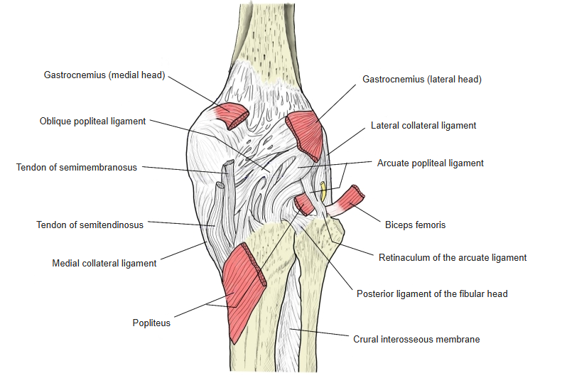
| 9 | Oblique popliteal ligament | Lateral condyle – tendon of the semimembranosus |
| 10 | Arcuate popliteal ligament | Lateral condyle – head of fibula |
The anterior and posterior cruciate ligaments together are sometimes called the cruciate ligaments of the knee.
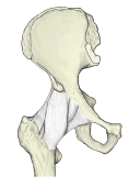
Hip joint ligaments (anterior) 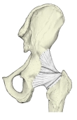
Hip joint ligaments (posterior) 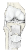
Knee joint (anterior) 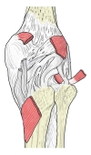
Knee joint (posterior)
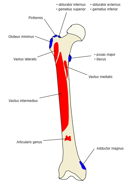
Muscle attachments
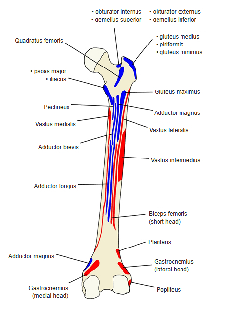
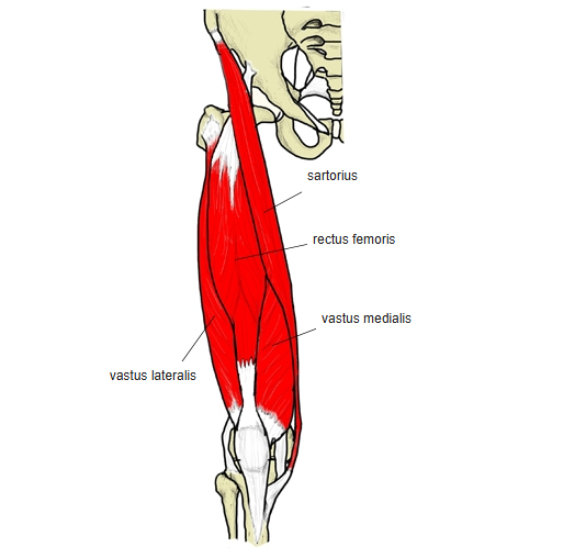
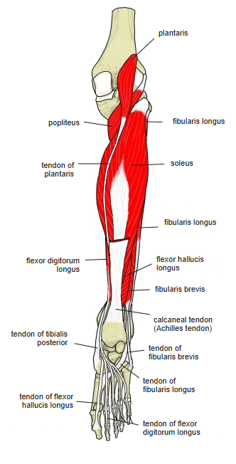
- Origin of: Vastus lateralis, Vastus intermedius, Vastus medialis, Articularis genus, Biceps femoris, Gastrocnemius, Plantaris, Popliteus
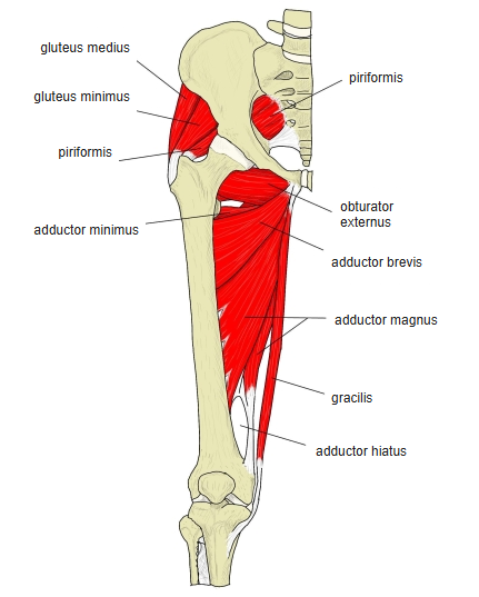
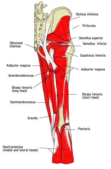
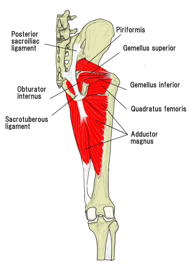
- Insertion of: Gluteus maximus, Gluteus medius, Gluteus minimus, Piriformis, Obturator internus, Gemellus superior, Gemellus inferior, Quadratus femoris, Obturator externus, Iliacus, Psoas major, Pectineus, Adductor longus, Adductor brevis, Adductor magnus
Origins: vastus lateralis, intermedius and medialis — the shaft (around the linea aspera and the front and sides); articularis genus — front of the lower shaft; biceps femoris (short head) — lateral lip of the linea aspera; gastrocnemius and plantaris — above the condyles; popliteus — lateral condyle. Insertions: gluteus medius, gluteus minimus, piriformis, obturator internus and the gemelli — greater trochanter and trochanteric fossa; quadratus femoris — quadrate tubercle; obturator externus — trochanteric fossa; iliacus and psoas major — lesser trochanter; gluteus maximus — gluteal tuberosity; pectineus — pectineal line; adductors — linea aspera (adductor magnus also the adductor tubercle).
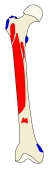
Muscle attachments (anterior) 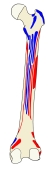
Muscle attachments (posterior) 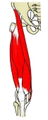
Muscles of the front of the thigh 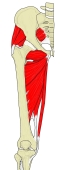
Hip and thigh, deep view 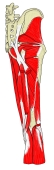
Hip and thigh, deep layer 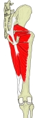
Deep hip muscles, posterior view 
Leg, posterior view (deeper)
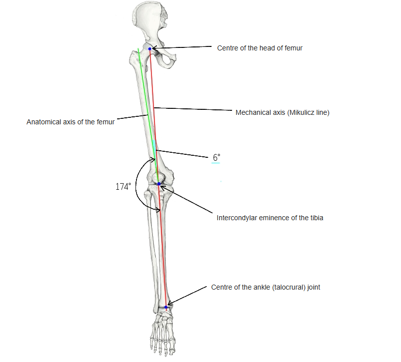
Axes of the lower limb
- With normal alignment, the large joints of the lower limb (hip, knee and ankle) lie on one straight line, the mechanical axis (Mikulicz line).
- The mechanical axis runs from the centre of the head of femur through the intercondylar eminence of the tibia to the centre of the ankle joint.
- In the tibia the anatomical and mechanical axes coincide; in the femur they differ by about 6°.
- So the anatomical axes of the femur and tibia are not in line: in the frontal plane they form an angle of about 174° opening laterally at the knee (the femorotibial angle).
Axes of the lower limb
Development
- Ossification of the femur begins at about the end of the 7th fetal week.
- The ossification centre of the distal epiphysis appears at the end of fetal life; its presence is used in forensic medicine to distinguish a full-term newborn from a fetus.
- The centre of the proximal epiphysis appears soon after birth; further centres appear in the greater and lesser trochanters and the medial and lateral condyles.
- The epiphyses fuse with the shaft at about 20–25 years.
References and sources
- Further reading: Femur, Wikipedia.