Hepatocolic ligament (Latin: ligamentum hepatocolicum)
The hepatocolic ligament is one of the peritoneal ligaments; it is stretched between the underside of the liver and the transverse colon (mainly near the right colic flexure).
Peritoneal ligament: a double fold of serous membrane that joins an organ to the wall or organs to one anotherOn the liver it is attached beside the gallbladder
Structures extending from the peritoneum into the abdominal cavity
- Mesentery : a double layer of peritoneum that attaches an organ to the parietal peritoneum of the abdominal wall and carries its blood vessels and nerves. Examples: the mesentery of the small intestine, the transverse mesocolon and others
- Peritoneal fold : a single layer raised into a ridge on the inner surface of the anterior abdominal wall by vessels or cord-like remnants. Examples: the median, medial and lateral umbilical folds
- Omentum : the peritoneum covering the stomach, extended from its borders upwards and downwards. Types: the lesser omentum (stomach to liver, a double layer) and the greater omentum (hangs down like a curtain; the double layer folds back on itself, so four layers fuse in the middle)
- Peritoneal ligament : a double layer that connects and supports an organ to the abdominal wall or to another organ. Examples: the falciform ligament, the gastrosplenic ligament
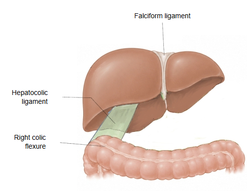
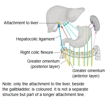
Its size varies greatly between individuals (variation), but on average it is about 5–6 cm long and wide.
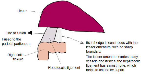
Right side: fused to the parietal peritoneum. Left side: continuous with the lesser omentum (its posterior layer).
There is no clear boundary between the hepatocolic ligament and the lesser omentum (posterior layer), but the lesser omentum carries many blood vessels and nerves while the hepatocolic ligament has almost none, so the two are not hard to tell apart.
Related terms
- Peritoneal ligaments
- Lesser omentum
- Hepatoduodenal ligament
- Hepatogastric ligament
- Liver
- Colon
- Transverse mesocolon
References and sources
- Further reading: Hepatocolic ligament, Wikipedia.
- Federative International Programme for Anatomical Terminology (FIPAT). Terminologia Anatomica, 2nd ed. (TA2), 2019.