Hyaloid canal (Latin: canalis hyaloideus)
Also called Cloquet's canal or Stilling's canal
Some sources say that the trace of the hyaloid artery disappears completely. Drawings of the eyeball are also divided: some show the hyaloid canal in the vitreous body and others leave it out.
In the embryo the hyaloid artery runs along this path, from about the sixth week until the seventh month of fetal life, and supplies the lens and the surrounding tissue with blood; it has largely disappeared by the eighth month, normally before birth.
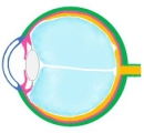 Section of the eyeball (1)
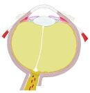 Section of the eyeball (2)
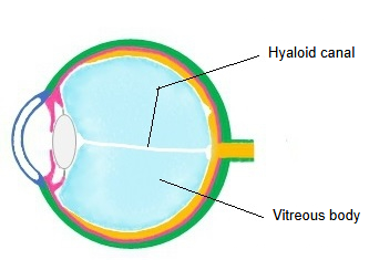
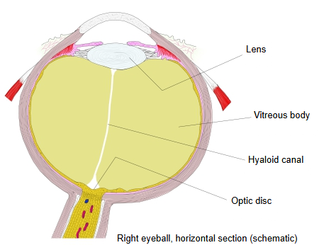
The hyaloid canal is a small, clear canal of thinner, more watery vitreous, about 1–2 mm wide. It begins at the optic disc and ends at the hyaloid fossa behind the lens; its course often sags downwards. It is formed by an infolding of the vitreous (hyaloid) membrane that encloses the vitreous body. In the fetus it contains the hyaloid artery, the continuation of the central retinal artery; after the artery regresses, the canal is filled with fluid. It has no known function in the adult eye. It does not take part in the changes of lens shape during accommodation, as was once thought: the volume of the lens hardly changes. Remnants of the artery may persist, for example as a small dot on the back of the lens or a tuft at the optic disc.
Related terms
References and sources
- Further reading: Hyaloid canal, Wikipedia.
- Further reading: Hyaloid artery, Wikipedia.
- Federative International Programme for Anatomical Terminology (FIPAT). Terminologia Anatomica, 2nd ed. (TA2), 2019.