Hyaloid fossa (Latin: fossa hyaloidea)
Also called the patellar fossa
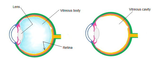
It lies at the front of the vitreous chamber, just behind the lens. The vitreous is firmly attached in a ring around the edge of the fossa; in the centre there is only a narrow potential space between the vitreous and the lens capsule, where blood or fluid can collect (the retrolental space). The hyaloid canal, the remnant of the fetal hyaloid artery, begins at the fossa and runs back through the vitreous towards the optic disc.
Reference websites
[Wikipedia]
· Hyaloid fossa, Wikipedia.
· Vitreous body, Wikipedia.
[Websites with illustrations]
· Shape of the lens and its position in the eye (figure) (BrainKart)
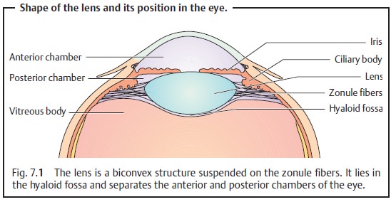
· Lens, chambers and hyaloid fossa (figure) (Ento Key)

· Eyeball in horizontal section (figure) (doctorlib.info)

Related terms
- Vitreous humour
- Vitreous chamber
- Vitreous membrane
- Zonular spaces
- Vitreous body
- Lens
- Eyeball
- Chambers of eyeball
- Ciliary zonule
- Posterior chamber of eyeball
References and sources
- Further reading: Hyaloid fossa, Wikipedia.
- Further reading: Vitreous body, Wikipedia.
- Federative International Programme for Anatomical Terminology (FIPAT). Terminologia Anatomica, 2nd ed. (TA2), 2019.