Mast cell (Latin: mastocytus)
Also called Mastocyte or the labrocyte
A cell of the immune system that triggers allergic reactions. Its name comes from the German Mast ('fattening'), but it has nothing to do with obesity.
In more detail
Mast cells develop from haematopoietic stem cells in the bone marrow. Their precursors travel in the blood and mature only after they settle in the tissues. They are found throughout the body in connective tissue, especially around small blood vessels and nerves: in the mucous membranes of organs such as the airways and the intestinal tract, and in the dermis of the skin. By some estimates the body holds about a trillion of them. Also called mastocytes.
The cell is round to oval with a central nucleus. Its cytoplasm is packed with coarse granules that stain strongly with basic dyes and change their colour (metachromasia), so the cell looks 'well-fed'; this is where the name comes from.
Functions
Mast cells are strongly linked with allergy, but allergic reactions are only one of their functions.
1. Triggering allergic reactions
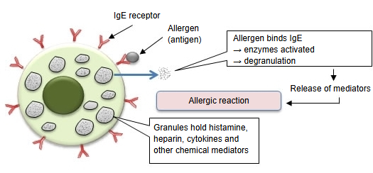
The cell surface carries many receptors for IgE antibodies (made by plasma cells derived from B lymphocytes). When an allergen binds to the IgE on these receptors, enzymes inside the cell are activated and the granules are released (degranulation). Chemical mediators in the granules, such as histamine and cytokines, are set free and cause the allergic reaction.
2. Defence of the mucous membranes and the skin against infection
Mediators released by mast cells widen the vessels and draw immune cells to the site, which helps the skin and mucous membranes fight bacteria and parasites. Mast cells resemble the basophils of the blood but are a separate cell line.
Chemical mediators released
| 1 | Histamine | Lowers blood pressure, makes vessels leaky, contracts smooth muscle, widens blood vessels and increases gland secretion. Basophils and some other cells also produce histamine. |
| 2 | Heparin | Prevents blood clotting. |
| 3 | Prostaglandins | Mainly prostaglandin D2: widens blood vessels and contracts bronchial smooth muscle. |
| 4 | Cytokines | Mainly IL-3, IL-4, IL-5, IL-6, IL-10, IL-13, I-309 (CCL1), GM-CSF, TNF-α and CXCL8 (IL-8). |
Types
The make-up of the proteins in the granules differs with the tissue in which the cells live.
| Where found | Granule proteins | |
|---|---|---|
| 1 | Connective tissue under the skin (hypodermis) and under the mucosa (connective tissue mast cells) | Tryptase and also large amounts of chymase |
| 2 | In the lungs and inside the mucosa, e.g. of the gut (mucosal mast cells) | Very little chymase |
Related terms
- Hematopoietic stem cell
- Fibrous connective tissue
- B lymphocyte
- Dermis
- Hypodermis (subcutaneous tissue)
- Skin
References and sources
- Further reading: Mast cell, Wikipedia.
- Federative International Programme for Anatomical Terminology (FIPAT). Terminologia Anatomica, 2nd ed. (TA2), 2019.