Latin: cutis
Overview
- The covering of the body surface, continuous with the mucous membranes at the lips, nostrils, eyelids, anus and urogenital openings.
- Thickness about 0.5–4 mm: thinnest on the eyelids, thickest on the back, palms and soles.
- Two main layers – the epidermis and the dermis – lie on the subcutaneous tissue.
- Functions: barrier and protection, temperature regulation (sweat and blood flow), sensation, vitamin D production.
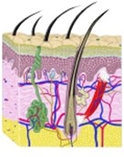
Skin, block diagram
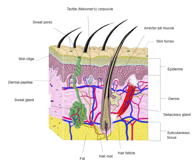
Layers
| Layer | Tissue | Contents |
|---|---|---|
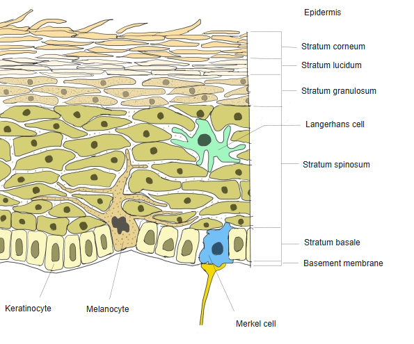
| Epidermis | Keratinized stratified squamous epithelium, no blood vessels | Keratinocytes, melanocytes, Langerhans cells, Merkel cells |
| Dermis | Dense connective tissue (collagen and elastic fibers) | Vessels, nerves and nerve endings, hair follicles, glands, arrector pili muscles |
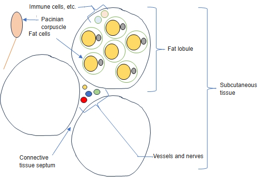
| Subcutaneous tissue | Loose connective tissue with fat lobules | Larger vessels and nerves, deep parts of glands and follicles, lamellar corpuscles |
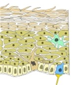
Layers of the epidermis 
Subcutaneous tissue (diagram)
Surface and types of skin
- Thick (glabrous) skin on the palms and soles: thick stratum corneum, no hair, many sweat glands; raised skin ridges form the fingerprints.
- Thin (hairy) skin covers the rest of the body; fine skin furrows divide it into small fields.
- Skin colour depends on melanin, carotene and the blood in the dermal vessels.
- Tension (cleavage) lines follow the main direction of the collagen fibers; incisions along them heal with narrower scars.
Skin appendages and nerve endings
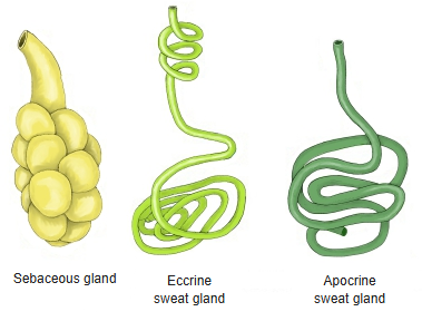
- Appendages: hair, nails, sweat glands, sebaceous glands and the mammary gland.
- Receptors: free nerve endings (pain, temperature), Merkel discs and tactile (Meissner) corpuscles (fine touch), lamellar (Pacinian) corpuscles (vibration and pressure), Ruffini endings (stretch).
Skin glands compared
References and sources
- Further reading: Skin, Wikipedia.