Posterior surface of radius (Latin: facies posterior radii)
Also called the dorsal surface
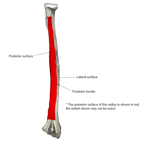
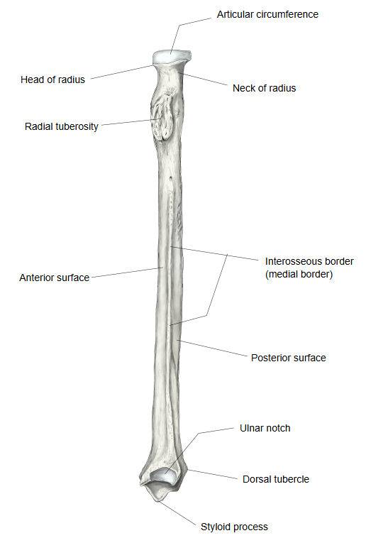
One of the three surfaces of the body (shaft) of the radius: the surface that faces backwards.
· It lies between the medial border (= interosseous border) and the posterior border.
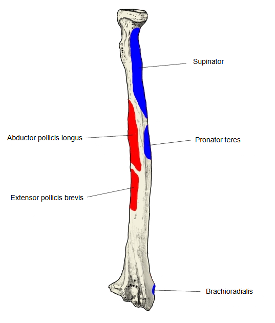
Upper third: rounded and smooth, covered by supinator.
Middle third: broad and slightly hollowed; it gives origin to abductor pollicis longus (above) and extensor pollicis brevis (below).
Lower third: broad and rounded, covered by the tendons that go on to run in the grooves for the extensor tendons on the back of the lower end, either side of the dorsal tubercle.
· It lies between the medial border (= interosseous border) and the posterior border.
Upper third: rounded and smooth, covered by supinator.
Middle third: broad and slightly hollowed; it gives origin to abductor pollicis longus (above) and extensor pollicis brevis (below).
Lower third: broad and rounded, covered by the tendons that go on to run in the grooves for the extensor tendons on the back of the lower end, either side of the dorsal tubercle.
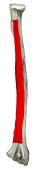 Right radius (posterior)
 Right radius (medial)
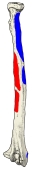 Muscle attachments (posterior)
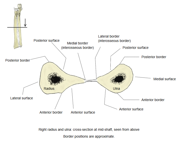 Cross-section of radius and ulna
· Video: The radius (the borders and surfaces are shown from about 4:10).
Where it is found
Related terms
- Anterior surface of radius
- Lateral surface of radius
- Posterior border of radius
- Interosseous border of radius
- Body of radius
- Dorsal tubercle of radius
- Grooves for extensor tendons
References and sources
- Further reading: Radius (bone), Wikipedia.
- Further reading: Abductor pollicis longus muscle, Wikipedia.
- Further reading: Extensor pollicis brevis muscle, Wikipedia.
- Federative International Programme for Anatomical Terminology (FIPAT). Terminologia Anatomica, 2nd ed. (TA2), 2019.