Grooves for extensor tendons (Latin: sulci tendinum musculorum extensorum)
Also called the grooves for extensor muscle tendons
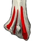 Right radius, distal end (posterior)
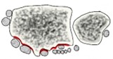 Cross-section at the wrist
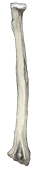 Right radius (posterior)
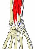 Forearm extensors (deep)
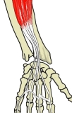 Extensor digitorum
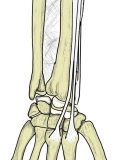 Radial wrist extensors
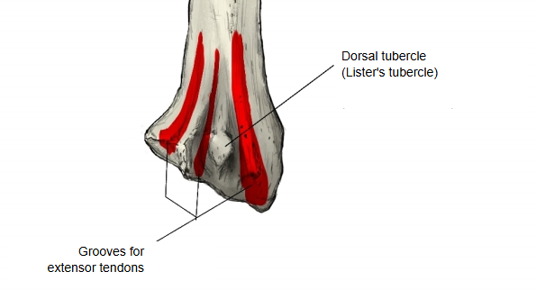
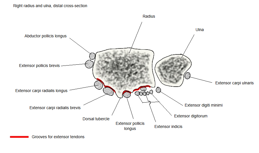
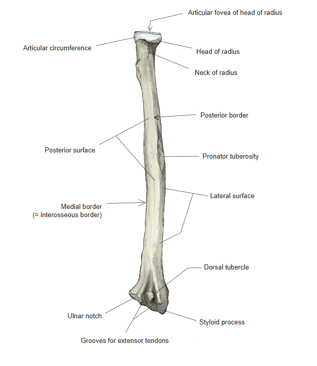
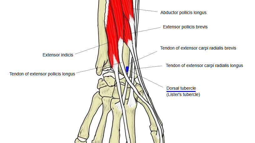
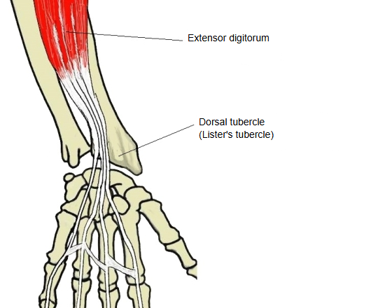
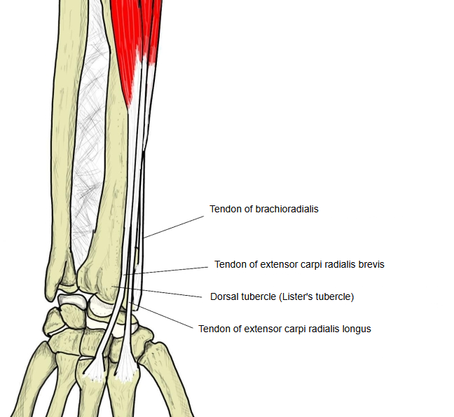
The back of the lower end of the radius is convex and gives attachment to the dorsal radiocarpal ligament. Classic descriptions count three grooves on it, from lateral to medial: a broad, shallow groove divided by a low ridge, for the tendons of extensor carpi radialis longus (lateral part) and extensor carpi radialis brevis (medial part); a narrow, deep groove running obliquely downwards and laterally, bounded laterally by a sharp ridge, for the tendon of extensor pollicis longus; and a broad groove for the tendons of extensor digitorum and extensor indicis.
for the
extensor
tendons
- Lateral groove (radial side) : the tendons of extensor carpi radialis longus and extensor carpi radialis brevis pass through it.
- Middle groove : the tendon of extensor pollicis longus passes through it; it lies just medial to the dorsal tubercle (Lister's tubercle), around which the tendon turns.
- Medial groove (ulnar side) : the tendons of extensor digitorum and extensor indicis pass through it.
· The ridges between the grooves give attachment to the extensor retinaculum, which turns the grooves into tunnels for the tendons and their synovial sheaths.
· Lateral to these grooves, the tendons of abductor pollicis longus and extensor pollicis brevis run in a groove on the lateral side of the styloid process. Extensor digiti minimi crosses the distal radioulnar joint, and extensor carpi ulnaris runs in a groove on the back of the ulna.
· Clinically the tunnels under the retinaculum are numbered as six dorsal compartments, 1 to 6 from lateral to medial; compartments 1 to 4 lie on the radius. Sources therefore differ in how many grooves they count on the radius: three in classic descriptions, four if the groove for abductor pollicis longus and extensor pollicis brevis is added.
Reference websites
[Wikipedia]
· Extensor retinaculum of the hand, Wikipedia.
· Radius (bone), Wikipedia.
[Websites with illustrations]
· Bones of the left forearm, posterior surface (Gray's Anatomy, fig. 214) (Wikimedia Commons)

· The radius (TeachMeAnatomy)
Where it is found
Related terms
- Dorsal tubercle of radius
- Posterior surface of radius
- Distal epiphysis of radius
- Styloid process of radius
- Ulnar notch
- Carpal articular surface
References and sources
- Further reading: Extensor retinaculum of the hand, Wikipedia.
- Federative International Programme for Anatomical Terminology (FIPAT). Terminologia Anatomica, 2nd ed. (TA2), 2019.