Stria vascularis (Latin: stria vascularis ductus cochlearis)
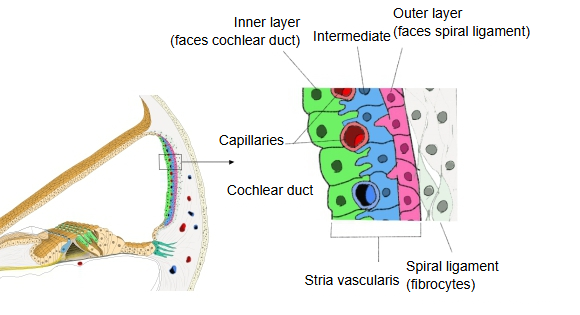
| 1 | Inner layer (marginal cells) | Faces the cochlear duct (inner side). Its cells transport potassium ions into the endolymph. |
| 2 | Intermediate layer (intermediate cells) | The layer in which the capillaries lie. |
| 3 | Outer layer (basal cells) | Faces the spiral ligament (outer side). Its cells are joined by gap junctions to the intermediate cells and to the connective-tissue cells (fibrocytes) of the spiral ligament. |
Note: Wikipedia names the layers after their cells (marginal cells, intermediate cells and basal cells); on this page they are called the inner, intermediate and outer layers.
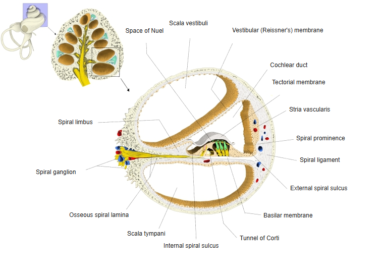
The stria vascularis runs along the whole length of the cochlea. It is unusual in containing capillaries within an epithelium; most epithelia have no blood vessels of their own. The marginal cells pump potassium ions into the endolymph, giving it a high potassium content and the positive endocochlear potential needed by the hair cells of the spiral organ.
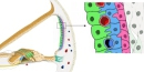 Three layers of the stria vascularis
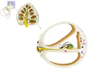 Cochlea and cochlear duct (section)
Reference websites
[Wikipedia]
· Stria vascularis of cochlear duct, Wikipedia.
[Websites with illustrations]
· Stria vascularis and its cell layers (Figure 1 of a research article) (Frontiers in Neurology)
· Section of the cochlear duct (image) (AMBOSS)

Related terms
- Cochlear duct
- Cochlea
- Spiral organ (organ of Corti)
- Membranous labyrinth
- Perilymphatic space
- Labyrinthine artery
References and sources
- Further reading: Stria vascularis of cochlear duct, Wikipedia.
- Federative International Programme for Anatomical Terminology (FIPAT). Terminologia Anatomica, 2nd ed. (TA2), 2019.