Superior extensor retinaculum (Latin: retinaculum musculorum extensorum superius)
Also called the transverse crural ligament
retinacula
of the leg
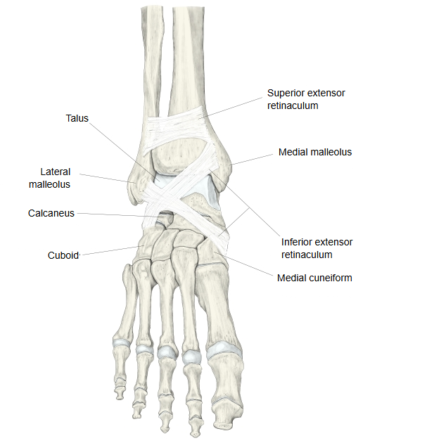
- Superior extensor retinaculum : stretched between the anterior borders of the tibia and the fibula.
- Inferior extensor retinaculum : Y-shaped, with a stem and two limbs.
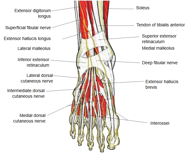
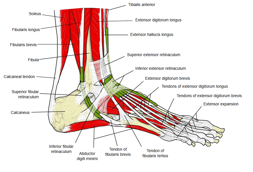
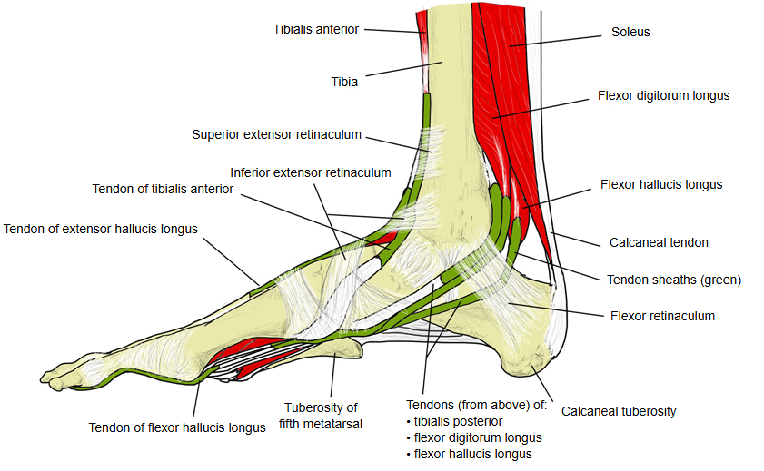
The structures that pass under the superior and inferior extensor retinacula of the leg are:
Others: anterior tibial artery, anterior tibial veins, deep fibular (peroneal) nerve.
· The back of the wrist also has an extensor retinaculum, but the upper limb has only one.
Under the superior extensor retinaculum the tendons lie, from medial to lateral, in this order: tibialis anterior, extensor hallucis longus, then extensor digitorum longus together with fibularis tertius; the anterior tibial vessels and the deep fibular nerve pass between extensor hallucis longus and extensor digitorum longus. Some texts describe three separate passages under it for these three groups; others describe separate compartments only under the inferior retinaculum. Usually only the tibialis anterior tendon has its own synovial sheath here. Above, the retinaculum is continuous with the deep fascia of the leg.
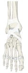 Extensor retinacula on the bones of the foot
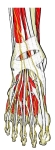 Dorsum of the foot
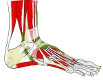 Ankle and foot, lateral view
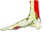 Ankle and foot, medial view
Reference websites
[Wikipedia]
· Superior extensor retinaculum of foot, Wikipedia.
· Inferior extensor retinaculum of foot, Wikipedia.
[Websites with illustrations]
· Tendon sheaths around the ankle, lateral view, with the transverse crural ligament (Gray's Anatomy, fig. 441) (Wikimedia Commons)

Related terms
- Inferior extensor retinaculum
- Tibialis anterior
- Extensor hallucis longus
- Extensor digitorum longus
- Fibularis tertius
- Tibia
- Fibula
References and sources
- Further reading: Superior extensor retinaculum of foot, Wikipedia.
- Federative International Programme for Anatomical Terminology (FIPAT). Terminologia Anatomica, 2nd ed. (TA2), 2019.