Latin: fibula · the lateral bone of the leg
Overview
- One of the two bones of the leg: a long bone on the lateral (little-toe) side, about as long as the tibia but much thinner.
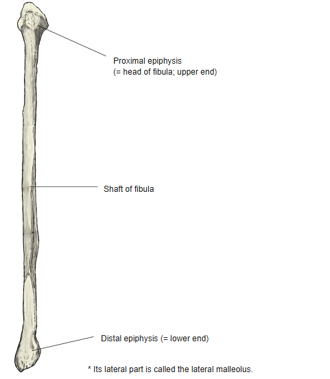
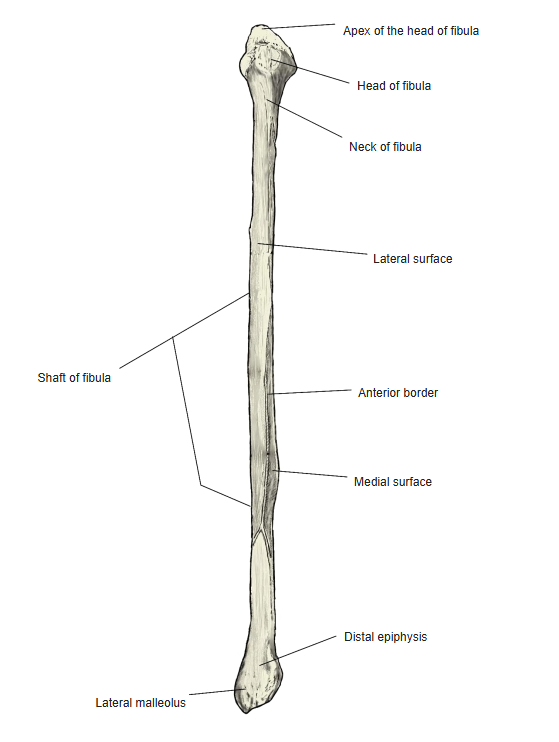
- It has an upper end (proximal epiphysis), a shaft and a lower end (distal epiphysis).
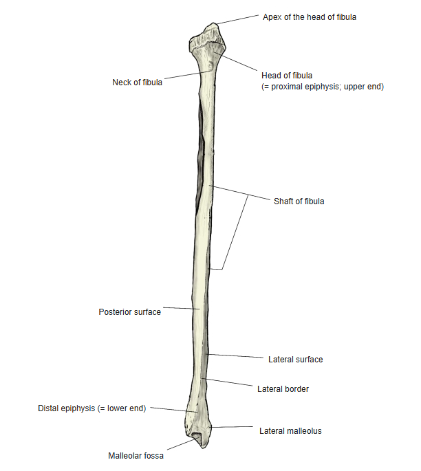
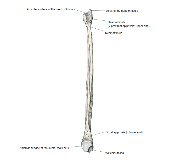
- Upper end: the head of fibula with its articular surface and apex, and the neck below it.
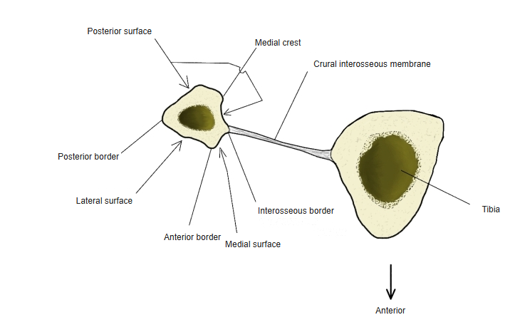
- Shaft: anterior, posterior and interosseous borders; medial, lateral and posterior surfaces, with the medial crest on the posterior surface.
- Lower end: the lateral malleolus, with its articular surface for the talus, the malleolar fossa behind it and the malleolar groove for the fibularis tendons.
- Most of the muscles attached to the fibula arise from the shaft; only a few attach to the head.
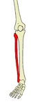
Bones of the lower limb 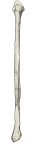
Parts of the fibula
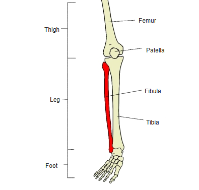
- Bones of the lower limb (one side)
- Pelvic girdle
- Hip bone ← ilium, ischium and pubis fused
- Free lower limb
- Thigh and knee
- Femur
- Patella
- Leg
- Tibia ← big-toe side
- Fibula ← little-toe side
- Foot
- Tarsal bones (7)
- Metatarsal bones (5)
- Phalanges (14)
- Thigh and knee
- Pelvic girdle
Illustrations

Right fibula (anterior) 
Right fibula (posterior) 
Right fibula (medial) 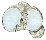
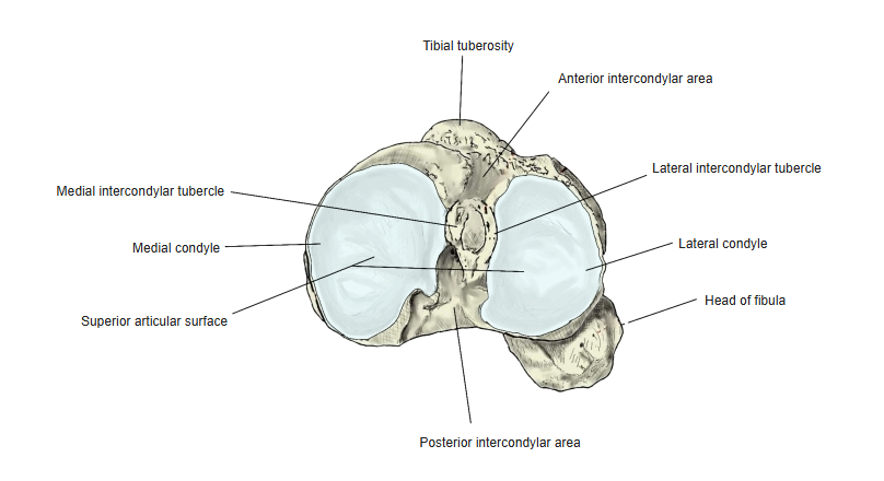
Tibia and fibula, from above 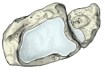
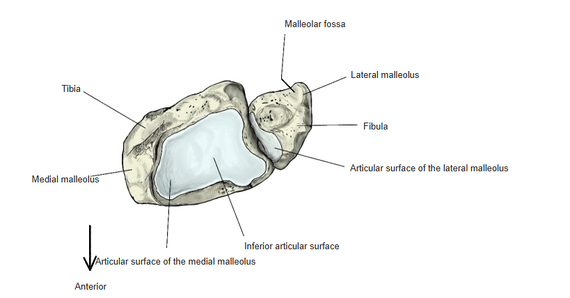
Tibia and fibula, from below 
Cross-section
Articulations
| # | Bone | Joint |
|---|---|---|
| 1 | Tibia | Tibiofibular joint |
| 2 | Tibia | Tibiofibular syndesmosis |
| 3 | Talus | Talocrural (ankle) joint |
Joints of the fibula
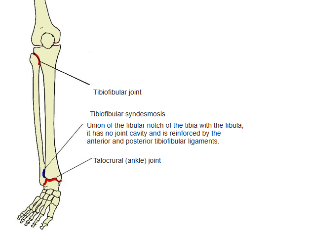
Tibiofibular joint: articular surface of the head of fibula ↔ lateral condyle of the tibia. Tibiofibular syndesmosis: lower end of the fibula ↔ fibular notch of the tibia (no joint cavity). Talocrural joint: articular surface of the lateral malleolus ↔ trochlea of the talus (with the inferior articular surface and medial malleolus of the tibia).
Ligaments
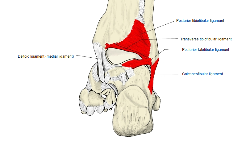
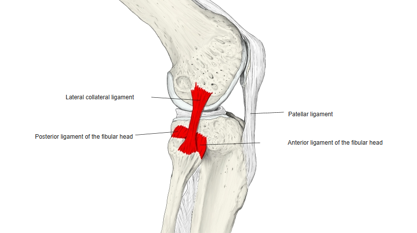
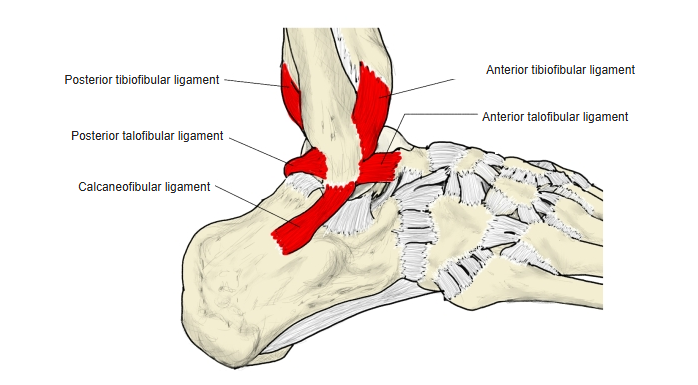
The following ligaments attach to the fibula. The list is intended to be comprehensive but may omit minor ligaments.
| # | Ligament | Connects |
|---|---|---|
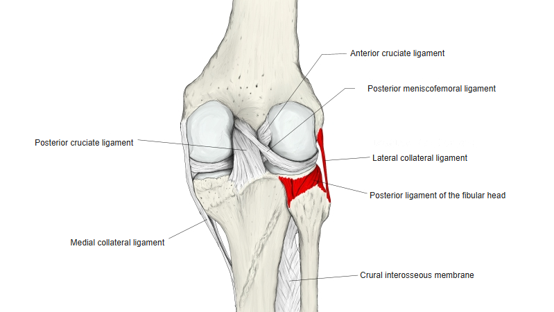
| 1 | Lateral (fibular) collateral ligament | Head of fibula – lateral epicondyle of femur |
| 2 | Anterior ligament of the fibular head | Head of fibula – lateral condyle of tibia |
| 3 | Posterior ligament of the fibular head | Head of fibula – lateral condyle of tibia |
| 4 | Anterior tibiofibular ligament | Lower end of fibula (lateral malleolus) – lower end of tibia |
| 5 | Posterior tibiofibular ligament | Lower end of fibula (lateral malleolus) – lower end of tibia |
| 6 | Transverse tibiofibular ligament | Lower end of fibula – lower end of tibia |
| 7 | Anterior talofibular ligament | Lateral malleolus – neck of talus |
| 8 | Posterior talofibular ligament | Malleolar fossa – lateral tubercle of talus |
| 9 | Calcaneofibular ligament | Lateral malleolus – calcaneus |
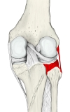
Right knee (posterior) 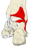
Right ankle (posterior) 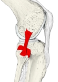
Right knee (lateral) 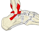
Right ankle (lateral)
Muscle attachments
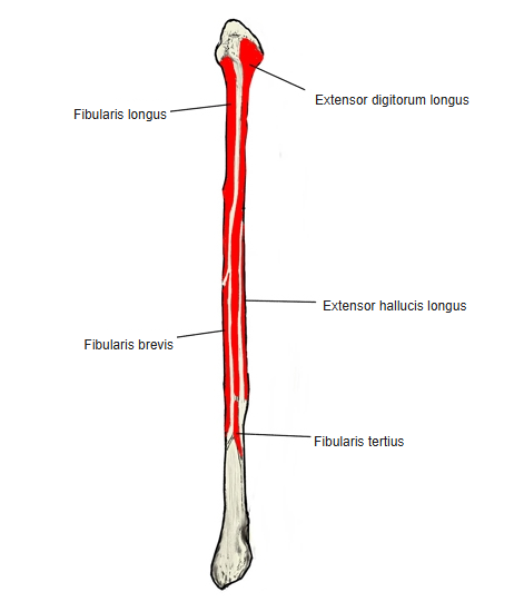
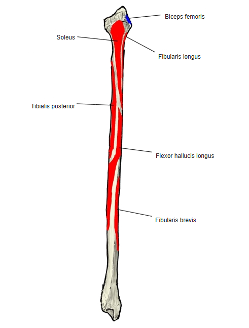
- Origin of: Tibialis posterior, Fibularis tertius, Extensor digitorum longus, Fibularis longus, Flexor hallucis longus, Extensor hallucis longus, Fibularis brevis, Soleus
- Insertion of: Biceps femoris
Origins: tibialis posterior and flexor hallucis longus — shaft, posterior surface; fibularis tertius and extensor hallucis longus — shaft, medial surface; extensor digitorum longus — anterior border; fibularis longus — mainly the lateral surface of the shaft (and the head); fibularis brevis — lateral surface; soleus — mainly the upper posterior part of the shaft and the head. Insertion: biceps femoris — head of fibula.

Muscle attachments (anterior) 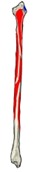
Muscle attachments (posterior)
Development
- The ossification centre of the shaft appears a little later than that of the tibia.
- The centre of the distal epiphysis appears at about 2 years, that of the proximal epiphysis at about 4 years.
- The shaft fuses with the epiphyses at about 21–22 years.
Blood supply
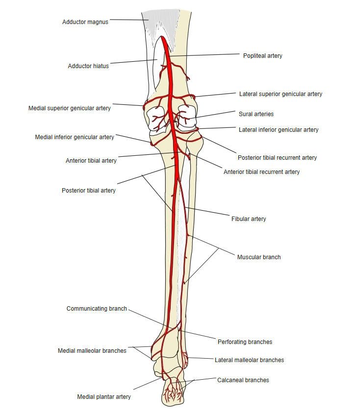
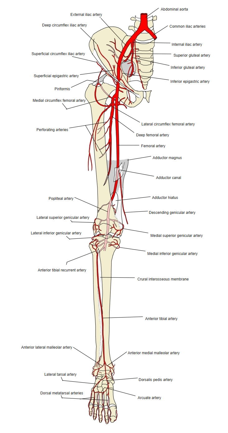
- The middle third of the shaft is supplied by a large nutrient vessel from the fibular artery; the periosteum also receives many small branches from the fibular artery.
- The head and the proximal epiphysis are supplied by a branch of the anterior tibial artery.
Arteries of the leg (posterior) 
Arteries of the lower limb (anterior)
References and sources
- Further reading: Fibula, Wikipedia.