Latin: tibia · also called the shinbone
Overview
- One of the two bones of the leg: a long bone on the medial (big-toe) side, much stronger than the fibula.
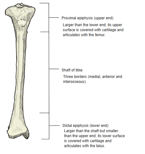
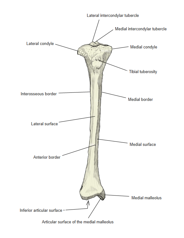
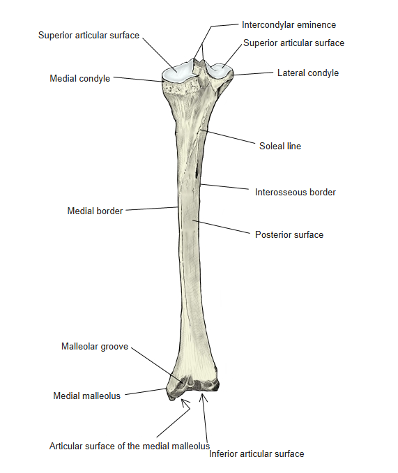
- It has an upper end (proximal epiphysis), a shaft and a lower end (distal epiphysis).
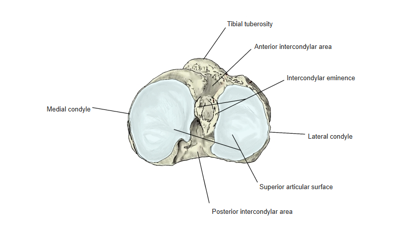
- Upper end: the medial and lateral condyles, whose superior articular surfaces articulate with the femur, separated by the intercondylar eminence (medial and lateral intercondylar tubercles) and the anterior and posterior intercondylar areas; the tibial tuberosity in front.
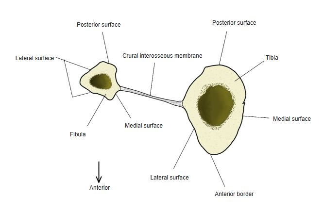
- Shaft: triangular in section, with 3 borders (anterior, medial, interosseous) and 3 surfaces (medial, lateral, posterior); the soleal line on the posterior surface.
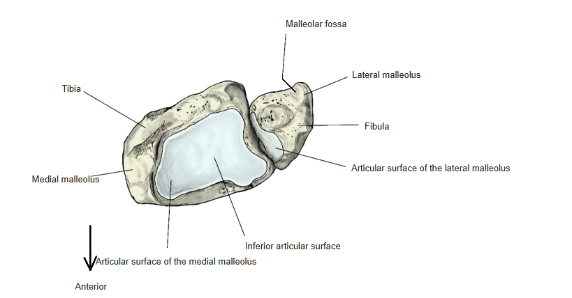
- Lower end: the medial malleolus with its articular surface and the malleolar groove, the inferior articular surface for the talus, and the fibular notch.
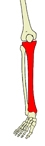
Bones of the lower limb 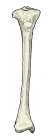
Parts of the tibia
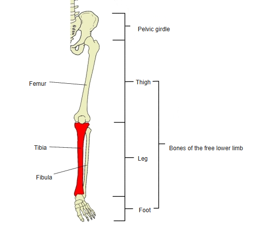
- Bones of the lower limb (one side)
- Pelvic girdle
- Hip bone ← ilium, ischium and pubis fused
- Free lower limb
- Thigh and knee
- Femur
- Patella
- Leg
- Tibia ← big-toe side
- Fibula ← little-toe side
- Foot
- Tarsal bones (7)
- Metatarsal bones (5)
- Phalanges (14)
- Thigh and knee
- Pelvic girdle
Illustrations
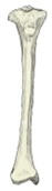
Right tibia (anterior) 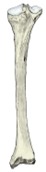
Right tibia (posterior) 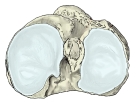
Upper end, seen from above 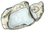
Lower end, seen from below 
Tibia and fibula (cross-section)
Articulations
| # | Bone | Joint |
|---|---|---|
| 1 | Femur | Knee joint |
| 2 | Fibula | Tibiofibular joint |
| 3 | Fibula | Tibiofibular syndesmosis |
| 4 | Talus | Talocrural (ankle) joint |
Joints of the tibia
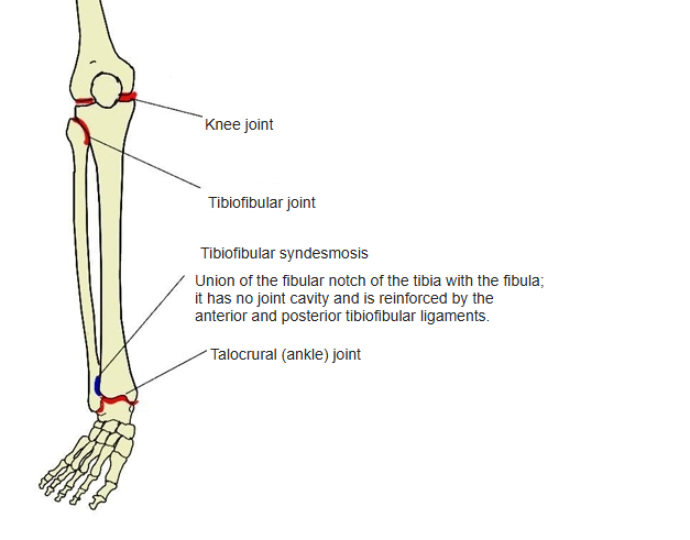
Knee joint: medial and lateral condyles of the tibia ↔ condyles of the femur. Tibiofibular joint: lateral condyle ↔ head of fibula. Tibiofibular syndesmosis: fibular notch ↔ lower end of the fibula (no joint cavity; reinforced by the anterior and posterior tibiofibular ligaments). Talocrural joint: inferior articular surface and medial malleolus ↔ trochlea of the talus; the lateral malleolus of the fibula also forms part of the mortise.
Ligaments
The following ligaments attach to the tibia. The list is intended to be comprehensive but may omit minor ligaments.
| # | Ligament | Connects |
|---|---|---|
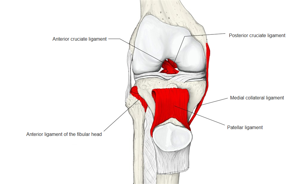
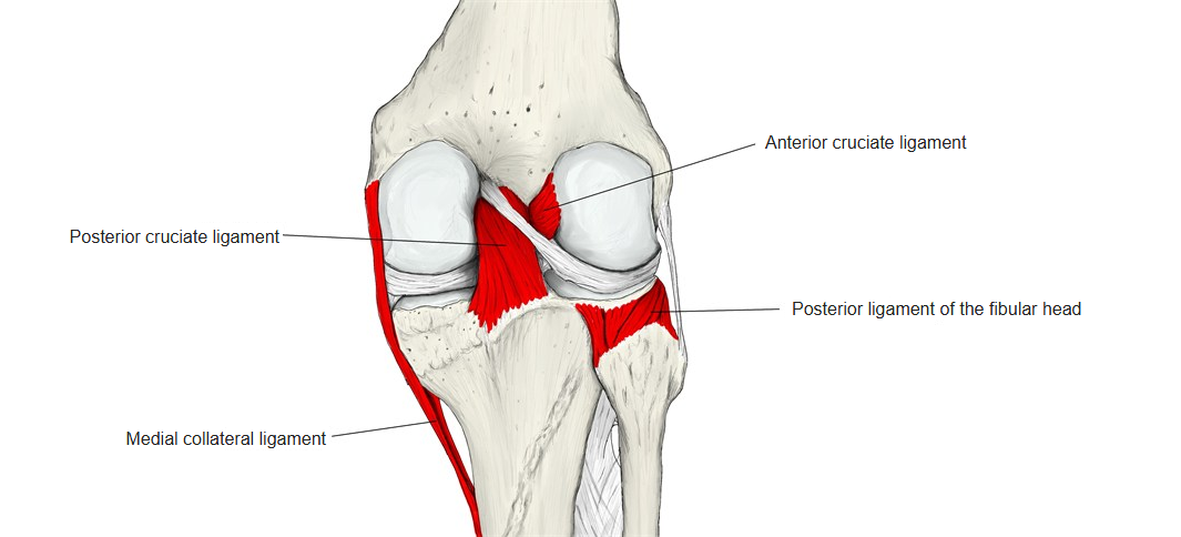
| 1 | Medial (tibial) collateral ligament | Medial condyle – medial epicondyle of femur |
| 2 | Patellar ligament | Tibial tuberosity – apex of patella |
| 3 | Anterior cruciate ligament | Anterior intercondylar area – lateral condyle of femur |
| 4 | Posterior cruciate ligament | Posterior intercondylar area – medial condyle of femur |
| 5 | Anterior ligament of the fibular head | Lateral condyle – head of fibula |
| 6 | Posterior ligament of the fibular head | Lateral condyle – head of fibula |
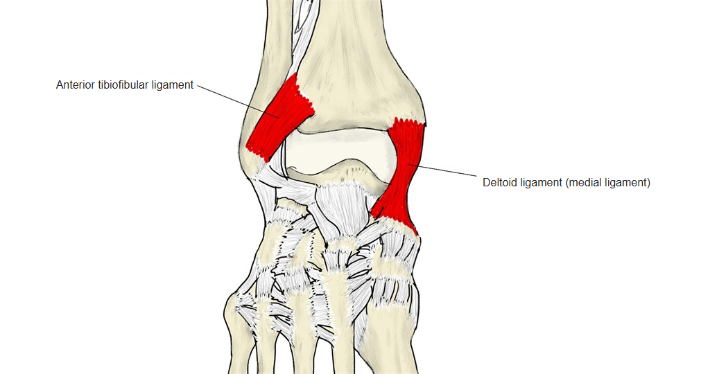
| 7 | Anterior tibiofibular ligament | Lower end of tibia – lower end of fibula (lateral malleolus) |
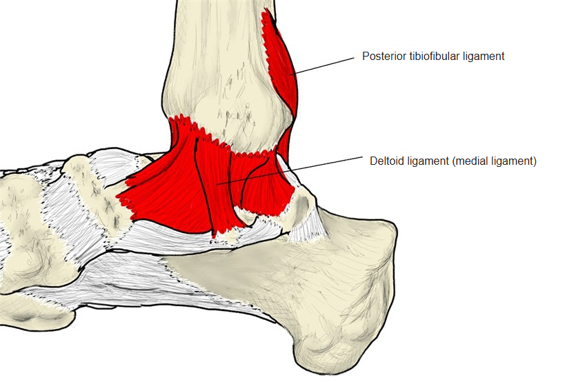
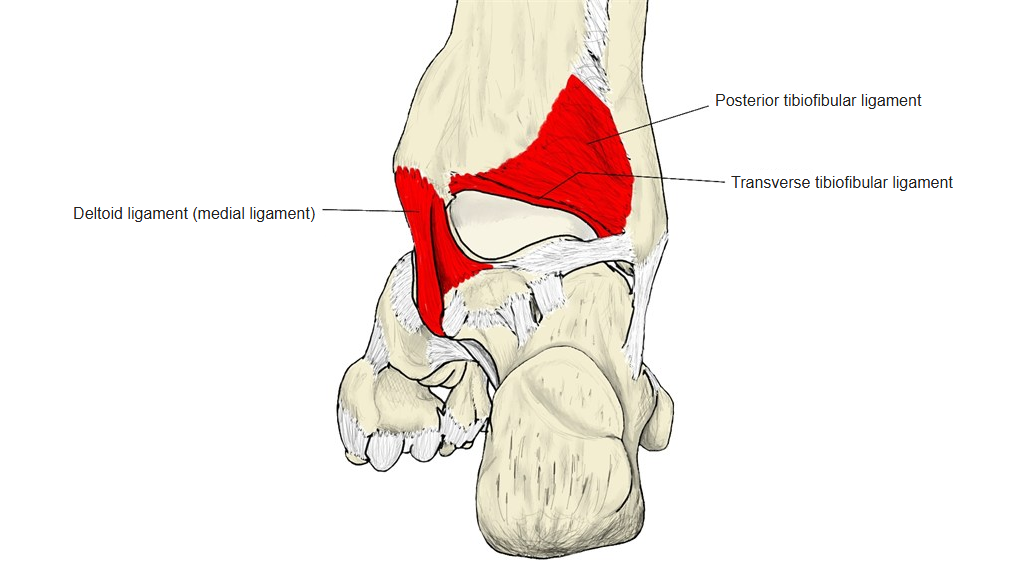
| 8 | Posterior tibiofibular ligament | Lower end of tibia – lower end of fibula (lateral malleolus) |
| 9 | Transverse tibiofibular ligament | Lower end of tibia – lower end of fibula |
| 10 | Deltoid (medial) ligament | Medial malleolus – navicular, calcaneus, talus |
The anterior and posterior cruciate ligaments together are sometimes called the cruciate ligaments of the knee.
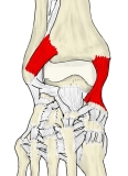
Right ankle (anterior) 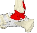
Right ankle (medial) 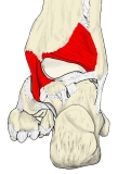
Right ankle (posterior) 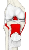
Right knee (anterior) 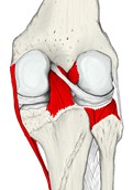
Right knee (posterior)
Muscle attachments
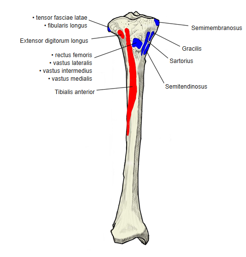
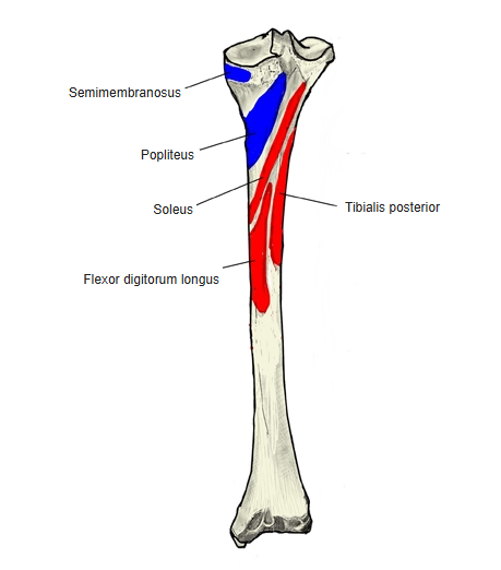
- Origin of: Tibialis posterior, Tibialis anterior, Flexor digitorum longus, Extensor digitorum longus, Fibularis longus, Soleus
- Insertion of: Vastus lateralis, Popliteus, Tensor fasciae latae, Rectus femoris, Vastus intermedius, Vastus medialis, Gracilis, Semitendinosus, Semimembranosus, Sartorius
Origins: tibialis posterior — shaft, posterior surface; tibialis anterior — lateral surface; flexor digitorum longus — posterior surface; extensor digitorum longus — lateral condyle and upper lateral surface; fibularis longus — lateral condyle; soleus — posterior surface (soleal line). Insertions: the four heads of the quadriceps femoris — tibial tuberosity through the patellar ligament; popliteus — posterior surface above the soleal line; tensor fasciae latae — lateral condyle through the iliotibial tract; gracilis, sartorius and semitendinosus — medial surface near the tibial tuberosity (pes anserinus); semimembranosus — medial condyle.
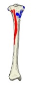
Muscle attachments (anterior) 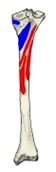
Muscle attachments (posterior)
Development and anomalies
- The ossification centre of the shaft appears in the 2nd fetal month; that of the proximal epiphysis at the end of fetal life or soon after birth; that of the distal epiphysis at about 2 years.
- A separate centre often appears in the tibial tuberosity at about 13 years.
- The lower epiphysis fuses with the shaft at about 16–19 years and the upper epiphysis at about 19–24 years (ages vary slightly between sources).
- Anomaly: the tibia may be markedly flattened from side to side, with an increased front-to-back diameter (platycnemia).
Blood supply
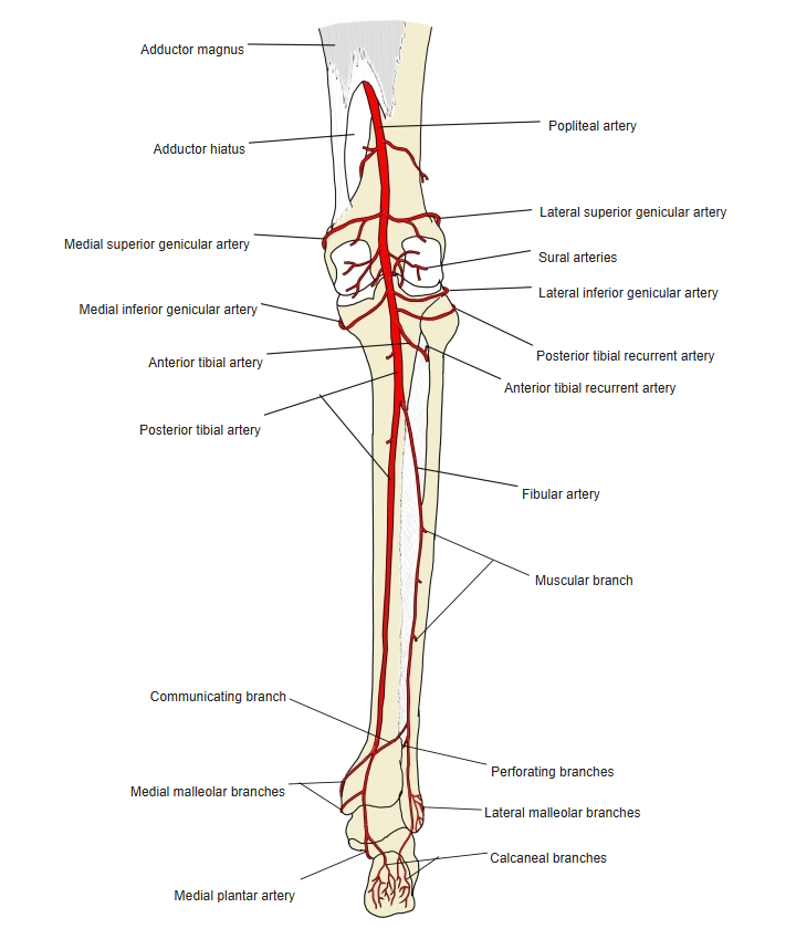
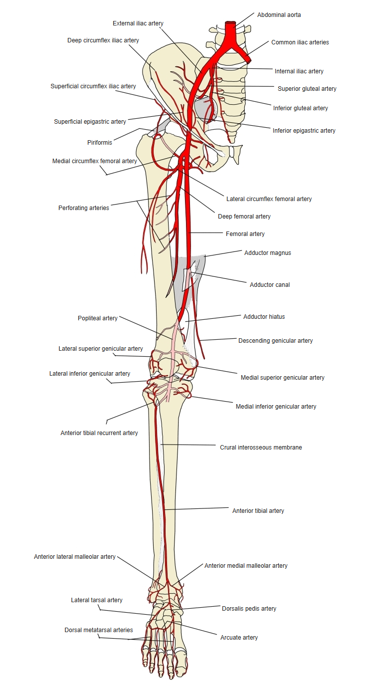
- Mainly from a nutrient artery (a branch of the posterior tibial artery), with periosteal vessels from the anterior tibial artery.
Arteries of the leg (posterior) 
Arteries of the lower limb (anterior)
References and sources
- Further reading: Tibia, Wikipedia.