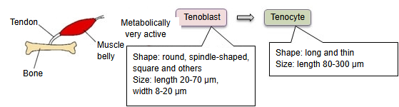
Tenoblast
Also called the tendon fibroblast
A cell found inside a tendon: the immature, actively working form of the tendon cell, which matures into a tenocyte.
A tendon contains three main kinds of cells: tenoblasts, tenocytes and chondrocytes (cartilage-like cells, found where the tendon is pressed against bone or attaches to it). Tenoblasts and tenocytes together make up about 90–95% of the cells in a tendon. "Tenoblast" is the name given inside the tendon to the fibroblasts found throughout the body's connective tissue, and tenoblasts change into tenocytes as they mature.
Tendon cells make and break down the collagen fibres and the rest of the extracellular matrix. Tenoblasts, which have more cytoplasm and organelles than tenocytes, do this much more actively; they are therefore more numerous in young tendons.