Chondrocyte (Latin: chondrocytus)
Also called Cartilage cell
Overview
· The share of the cartilage taken up by chondrocytes depends on the type of cartilage, but it is at most about 10%.
· Cartilage has no blood vessels, nerves or lymph vessels, so chondrocytes are nourished by diffusion through the matrix from the perichondrium or, in joints, from the synovial fluid. They arise from chondroblasts, the young cells of the inner layer of the perichondrium.
It is lowest in fibrocartilage, where thick bundles of collagen fibres fill most of the matrix; hyaline and elastic cartilage have more cells.
Figures for the share of cells in each type differ considerably between sources.
of
cartilage
- Cartilage cells : chondrocytes: secrete the cartilage matrix (everything except the water) and maintain the function of cartilage; only a small part of its volume.
- Cartilage matrix : = extracellular matrix
- Fibrous component : mainly type II collagen (hyaline cartilage); also type I collagen (fibrocartilage) and elastic fibres (elastic cartilage).
- Ground substance : amorphous: proteoglycans (most of it), hyaluronan and glycoproteins.
- Water : most of the matrix, held by the proteoglycans; important for the diffusion of nutrients and wastes.
· A chondrocyte normally has one nucleus, but cells with two nuclei are seen from time to time.
chondrocytes
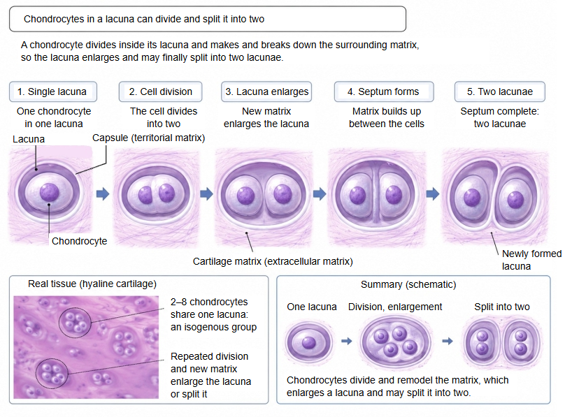
in a lacuna
- One cell : the cell stays single and does its work to the end.
- Several cells
- Stay together : a single cell divides, and the daughter cells do their work together in the one lacuna.
- Split apart : a single cell divides, the lacuna itself enlarges, a partition forms, and it divides into several lacunae.
A chondrocyte can divide inside its lacuna. The daughter cells may stay together as an isogenous group (cell nest) of a few cells. As they lay down new matrix the lacuna enlarges, and a septum of matrix may form between the cells and split it into two or more lacunae. This growth from within the cartilage is interstitial growth; growth by new cells from the perichondrium is appositional growth.
Size and shape
The shape of chondrocytes differs with the type of cartilage, and even within one type with the place where they lie.
| Type of cartilage and site | Superficial cells: shape and arrangement | Deep cells: shape and arrangement | Why (functional meaning) | |
|---|---|---|---|---|
| 1 | Articular cartilage (hyaline cartilage without a perichondrium) | Flat, lying side by side parallel to the surface | Rounded; deeper still they line up in vertical columns | The flat surface cells let the joint surface take strong friction (shear), and the round and column cells deeper down bear heavy loads (vertical compression). |
| 2 | Other hyaline cartilage (trachea, costal cartilage etc.) | Somewhat flat to spindle-shaped (still chondroblasts) | Large, round or oval (forming isogenous groups) | Young cells newly formed from the perichondrium at the surface grow into mature round cells as they move deeper and secrete matrix. |
| 3 | Elastic cartilage (auricle, epiglottis etc.) | Smallish, round or oval (fairly dense) | Large, round (very densely packed) | The whole tissue is laced with a network of elastic fibres, so the cells keep their basic round shape from the surface to the depth and become more densely packed deeper in. |
| 4 | Fibrocartilage (intervertebral disc, menisci etc.) | In rows along the fibres (may look rather flat) | In rows along the fibres (scattered in single file) | Thick bundles of type I collagen fibres run through it from surface to depth, so the cells are pressed into the gaps between the bundles and can only line up in rows. |
Secretions
The main substances secreted by chondrocytes are the following; their proportions differ with the type of cartilage.
· Hyaline cartilage: secretes mainly type II collagen fibres and proteoglycans.
· Elastic cartilage: also secretes much elastin (elastic fibres).
· Fibrocartilage: secretes mainly type I collagen fibres.
| Kind of substance | Name and role | ||
| 1 | Proteins | Glycoproteins and proteoglycans | · Collagen fibres (= collagen fibres, mainly type II): give the tissue a strong framework, with strength and stiffness. · Proteoglycans (such as aggrecan): carry large amounts of polysaccharide chains, hold much water and act as a cushion. |
| Simple protein | · Elastin (elastic fibres): gives the tissue supple elasticity and the ability to stretch. | ||
| 2 | Carbohydrates (polysaccharides) | · Hyaluronan (hyaluronic acid, one of the glycosaminoglycans): holds very large amounts of water and gives the tissue shock absorption and lubrication. | |
Distribution
How chondrocytes are distributed in cartilage differs with the type of cartilage.
In hyaline cartilage and some other types it also differs with the site.
| Feature | Hyaline cartilage | Elastic cartilage | Fibrocartilage | |
|---|---|---|---|---|
| 1 | Cell density | Moderate | Rather high | Low |
| 2 | Arrangement | Scattered irregularly (regular in articular cartilage and epiphyseal cartilage) | Scattered irregularly | In rows along the fibre bundles |
| 3 | Single cells | Many | Many | Few |
| 4 | Cell groups (cell nests, isogenous groups) | Common | Fairly common | Few |
| 5 | Size of the lacunae | Rather large | Rather large | Smaller |
| 6 | Distance between cells | Moderate | Rather small | Large (much collagen) |
| 7 | Characteristic | Typical cartilage, with round cells scattered in the matrix | Cells scattered in a network of elastic fibres | Cells in single file between bundles of type I collagen |
Related terms
- Cartilage
- Cartilage lacuna
- Cartilage matrix
- Perichondrium
- Hyaline cartilage
- Elastic cartilage
- Articular cartilage
References and sources
- Further reading: Chondrocyte, Wikipedia.
- Federative International Programme for Anatomical Terminology (FIPAT). Terminologia Anatomica, 2nd ed. (TA2), 2019.