Tendon sheath (Latin: vagina tendinis)
The tube-like covering that encloses a tendon in the hand, the foot and similar places, where the tendon runs over bone or under a retinaculum; it lets the tendon glide smoothly.
More detail
Structure
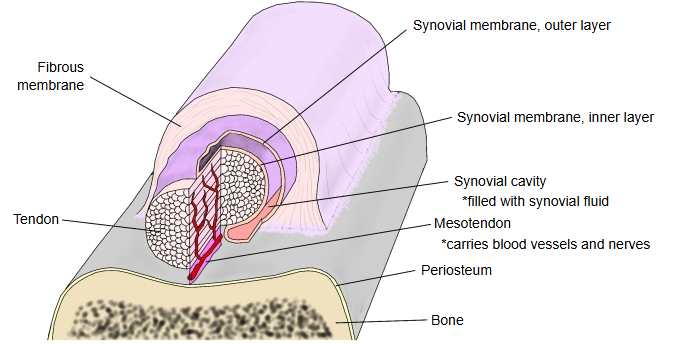
A tendon sheath has two parts: an outer, tunnel-like fibrous membrane (fibrous sheath) and an inner, sac-like synovial membrane (synovial sheath).
- Fibrous membrane : the outer layer: a tunnel of dense connective tissue
- Synovial membrane : a sac of two layers; the space inside it (the synovial cavity) is filled with synovial fluid
- Inner (visceral) layer : closely attached to the tendon
- Outer (parietal) layer : joined to the fibrous membrane
- Mesotendon : the fold joining the two layers; in places (as in the fingers) it is called the short and long vincula. It is the path of the blood vessels and nerves to the tendon.
Note on the name
Usage varies. Some texts head the section "synovial sheaths and fibrous sheaths (tendon sheaths)", so that "tendon sheath" may mean the fibrous sheath only; others say that the synovial sheath is also called the tendon sheath; and others use the name for the whole structure. Sources differ.
In the figure above, the fibrous membrane corresponds to the fibrous sheath and the synovial membrane to the synovial sheath; here "tendon sheath" is taken to mean the two together.
Where they are found
Hand
On the back of the hand the sheaths run from under the extensor retinaculum onto the dorsum of the hand; they are generally smaller than those on the palmar side. On both sides each sheath carries the tendons of the muscle of the same name.
| Dorsal | 1 | Sheath of abductor pollicis longus and extensor pollicis brevis | |
| 2 | Sheath of the radial wrist extensors (extensor carpi radialis longus and brevis) | ||
| 3 | Sheath of extensor pollicis longus | communicates with the sheath of the radial wrist extensors | |
| 4 | Sheath of extensor digitorum and extensor indicis | ||
| 5 | Sheath of extensor digiti minimi | ||
| 6 | Sheath of extensor carpi ulnaris | ||
| Other | 7 | Sheath of flexor carpi radialis | on the palmar side: around the tendon where it passes the scaphoid and runs in its own groove on the trapezium to its insertion |
| 8 | Bursa of extensor carpi radialis brevis | a small synovial bursa under its tendon near its insertion | |
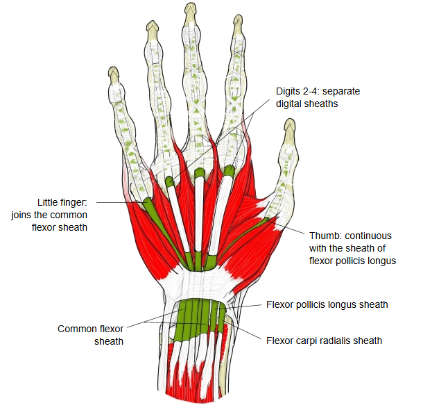
| Palmar | 1 | Sheath of flexor pollicis longus | |
| 2 | Common flexor sheath | for the tendons of flexor digitorum superficialis and flexor digitorum profundus, through the carpal tunnel under the flexor retinaculum | |
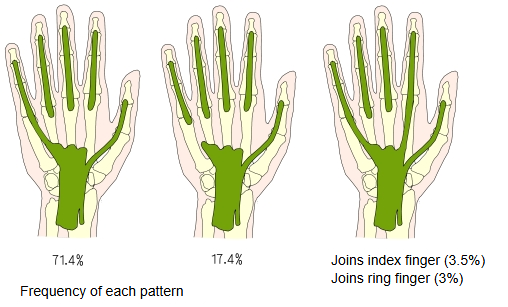
| 3 | Digital sheaths of the fingers | for the superficial and deep finger flexors. Those of the 2nd–4th fingers are separate, running from about the metacarpal heads to the distal phalanges; that of the thumb continues from the sheath of flexor pollicis longus, and that of the little finger usually joins the common flexor sheath. |
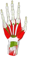 Palmar sheaths of the hand
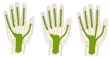 Patterns of the palmar sheaths
Foot
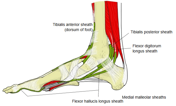
| Medial malleolar region | 1 | Sheath of tibialis posterior | the highest; it lies in front of and above the sheath of flexor digitorum longus |
| 2 | Sheath of flexor digitorum longus | between the sheaths of tibialis posterior and flexor hallucis longus; in the sole it ends near where its tendon crosses that of flexor hallucis longus | |
| 3 | Sheath of flexor hallucis longus | the lowest; it runs behind and below the tendon of flexor digitorum longus towards the base of the big toe | |
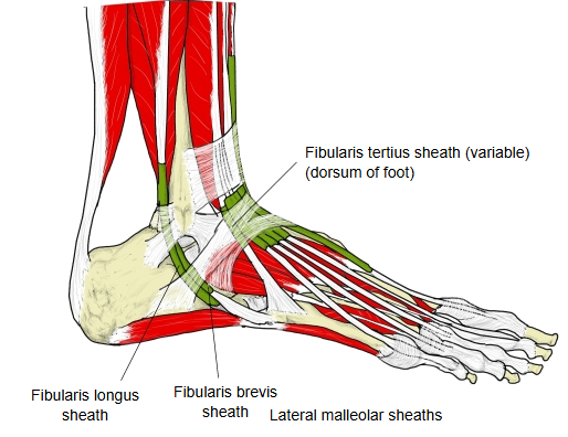
| Lateral malleolar region | 1 | Common sheath of the fibular muscles | carries the tendons of both fibularis longus and fibularis brevis behind the lateral malleolus; below it, it divides into a separate sheath for each tendon (some atlases draw the two sheaths as separate) |
| 2 | Plantar sheath of fibularis longus | in the sole, around the tendon as it runs forwards and medially across the foot in the groove of the cuboid, under the long plantar ligament | |
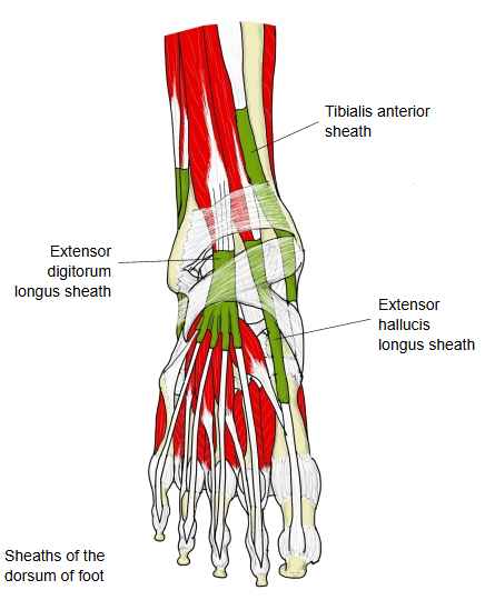
| Dorsum of the foot | 1 | Sheath of tibialis anterior | the most medial |
| 2 | Sheath of extensor hallucis longus | in the middle | |
| 3 | Sheath of extensor digitorum longus | the most lateral; it also holds the tendon of fibularis tertius | |
| Digital sheaths of the toes | In the sole, the flexor tendons of each toe are enclosed in a common synovial sheath on the plantar side of the toe: that of flexor hallucis longus for the big toe, and those of flexor digitorum longus and flexor digitorum brevis for the 2nd–5th toes. Their structure is like that of the fingers, but they do not communicate with the sheaths at the ankle. | ||
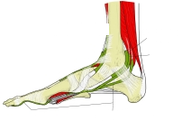 Sheaths at the medial ankle
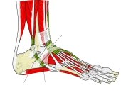 Sheaths at the lateral ankle
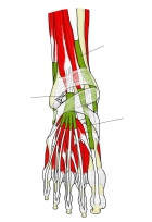 Sheaths on the dorsum of the foot
Related terms
- Synovial membrane
- Flexor retinaculum (hand)
- Carpal tunnel
- Flexor pollicis longus
- Flexor digitorum superficialis
- Tibialis posterior
- Fibularis longus
References and sources
- Further reading: Tendon sheath, Wikipedia.
- Federative International Programme for Anatomical Terminology (FIPAT). Terminologia Anatomica, 2nd ed. (TA2), 2019.