Pectinate ligament of iridocorneal angle
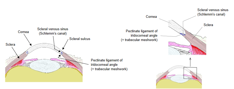
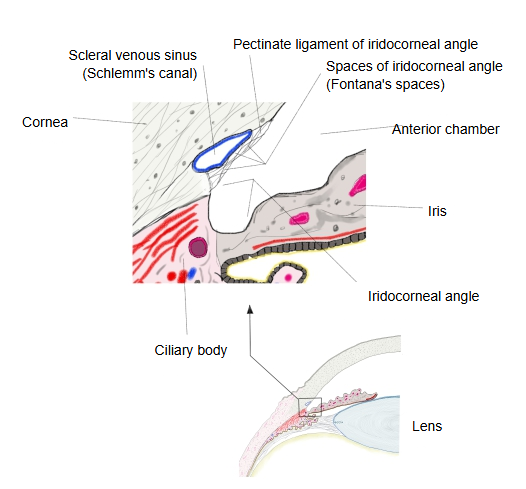
In current terminology this tissue is called the trabecular meshwork (Latin: reticulum trabeculare). Most of the aqueous humour leaves the eye through it.
The name trabecular meshwork is preferred because it describes the strands of the pectinate ligament of the iridocorneal angle, and the spaces of the iridocorneal angle between them, more accurately. Its inner, posterior part, which passes into the stroma of the iris and the ciliary body, is the uveal part (named after the uvea); its outer, anterior part, which runs from the inner side of the scleral spur forwards to the edge of the corneal endothelium, is the corneoscleral part. Next to the sinus lies a thin layer of juxtacanalicular tissue, through which the aqueous humour passes into the scleral venous sinus; reduced outflow here raises the pressure in the eye (glaucoma).
Reference websites
[Wikipedia]
· Trabecular meshwork, Wikipedia.
[Websites with illustrations]
· Cornea, sclera, ciliary body and iris at the angle (illustration) (image on cloudfront.net)

· Angle of the anterior chamber (illustration) (image on cloudfront.net)

Related terms
- Iridocorneal angle
- Spaces of iridocorneal angle
- Scleral venous sinus
- Scleral sulcus
- Cornea
- Corneal endothelium
- Sclera
- Iris
- Stroma of iris
- Ciliary body
- Uvea
- Anterior chamber of eyeball
- Aqueous humor
References and sources
- Further reading: Trabecular meshwork, Wikipedia.
- Federative International Programme for Anatomical Terminology (FIPAT). Terminologia Anatomica, 2nd ed. (TA2), 2019 (trabecular meshwork, with its corneoscleral and uveal parts).