Latin: musculus abductor hallucis
Overview
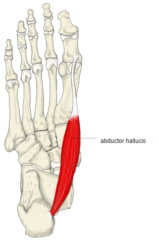
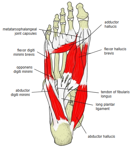
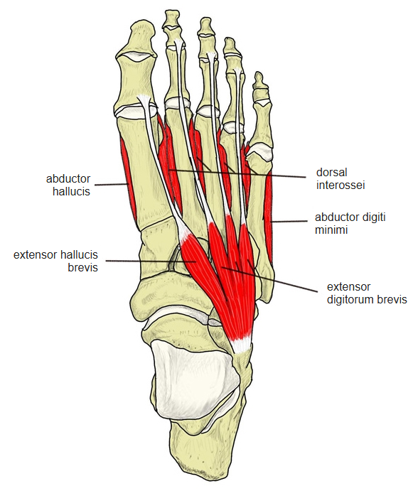
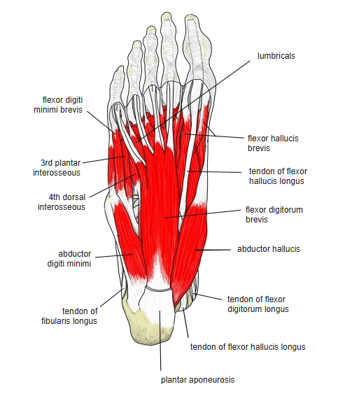
- Lies along the medial border of the foot and forms the bulge there; thick behind, flattening forwards.
- Ends in a common tendon with the medial head of the flexor hallucis brevis.
- With weight on the foot, helps to hold up the medial longitudinal arch.
Position in the classification
Lower Limb › Foot › Sole of foot › Muscles of the big toe
- Abductor hallucis
- Flexor hallucis brevis
- Adductor hallucis
Illustrations
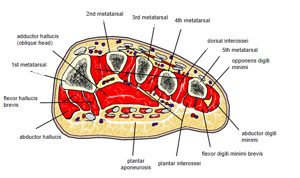
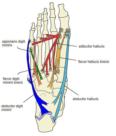
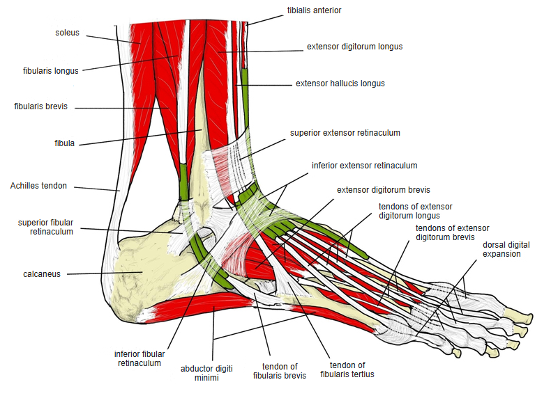
Origin / insertion
- Origin: medial process of the calcaneal tuberosity, superficial layer of the flexor retinaculum, plantar aponeurosis
- Insertion: medial side of the base of the proximal phalanx of the big toe and its medial sesamoid bone
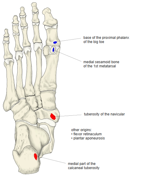
Action
- Abducts and flexes the big toe at the metatarsophalangeal joint
Innervation
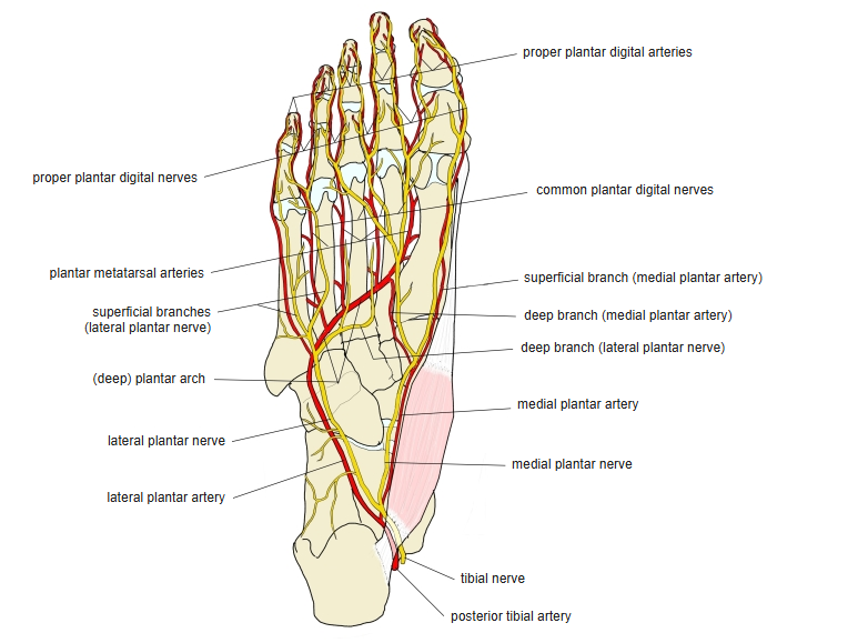
- Medial plantar nerve (S1–S2)
Blood supply
- Medial plantar artery
Related terms
- Flexor hallucis brevis
- Adductor hallucis
- Plantar aponeurosis
- Calcaneus
- Sesamoid bone
- Medial plantar nerve
References and sources
- Further reading: Abductor hallucis muscle, Wikipedia.