Latin: musculus deltoideus
Overview
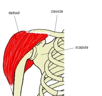
- One of the six muscles of the shoulder girdle (the scapulohumeral muscles); the thick muscle that gives the shoulder its rounded contour.
- Named after the Greek capital letter delta (Δ) because of its triangular shape.
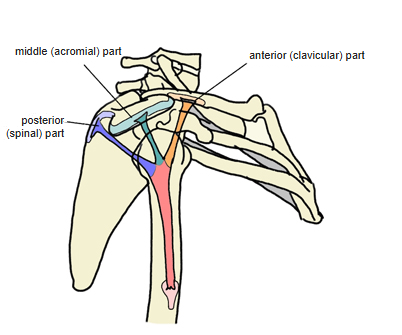
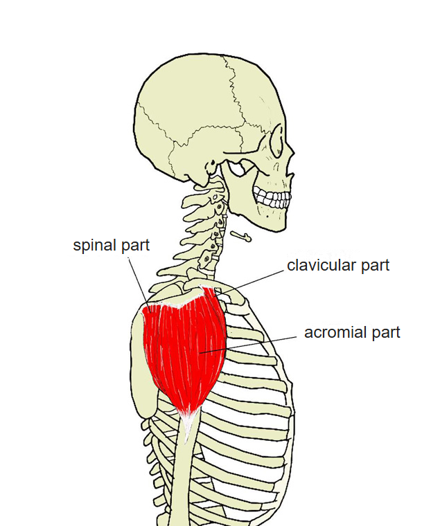
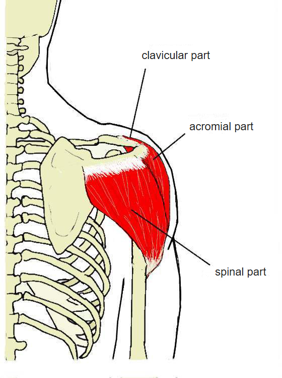
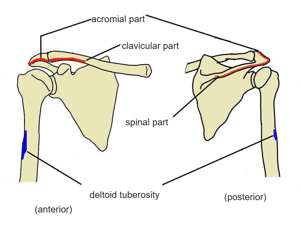
- Divided by origin into three parts: clavicular (anterior), acromial (middle) and spinal (posterior).
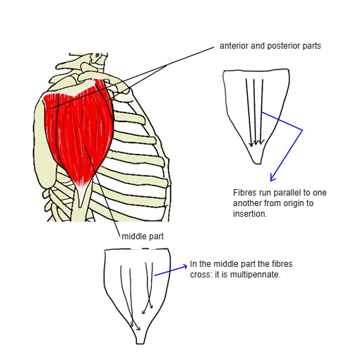
- In the anterior and posterior parts the fibres run parallel from origin to insertion; in the middle (acromial) part the fibres are multipennate.
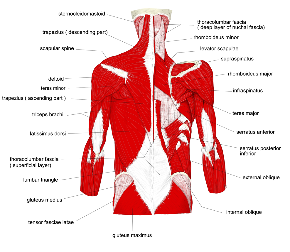
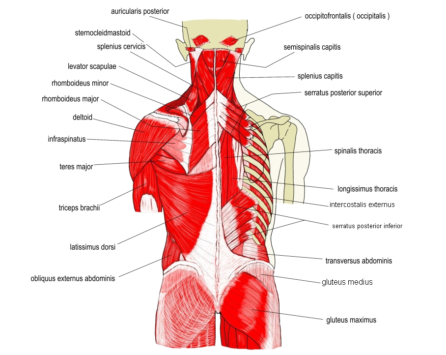
- Continuous in places with the trapezius, brachialis, pectoralis major and infraspinatus.
- Electromyography suggests at least seven functional units that the nervous system can control separately.
- Average mass about 192 g in a study of 30 shoulders (range about 84–366 g).
Tendons
- The origin tendons are cord-like and the insertion tendons sheet-like; both are seen mainly on the deep (bone) side.
- There are often 4 origin tendons and 5 insertion tendons; near its origin the posterior part consists of a thin membranous tendon only.
Deltopectoral groove and triangle
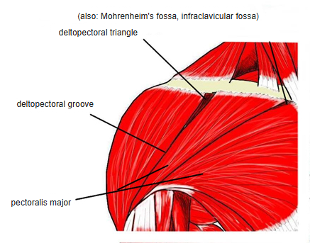
- Deltopectoral groove: the furrow between the deltoid and the pectoralis major; the cephalic vein runs in it.
- Deltopectoral triangle (Mohrenheim's fossa, infraclavicular fossa): the triangular hollow at the upper end of the groove, just below the clavicle.
Position in the classification
Upper Limb › Shoulder girdle
Illustrations
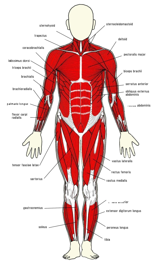
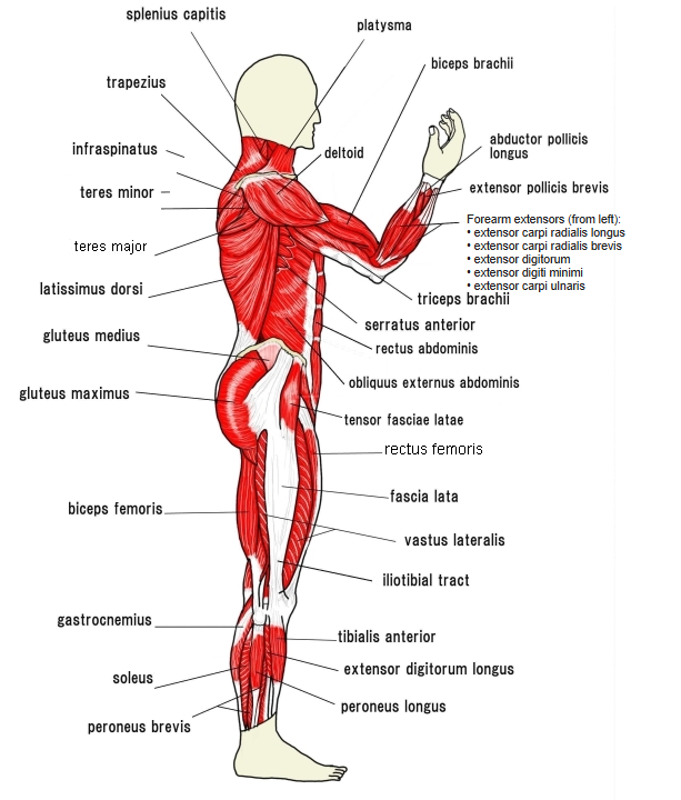
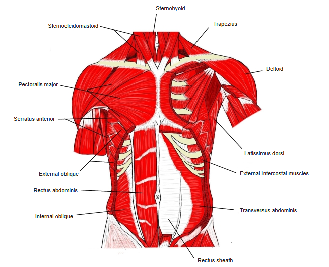
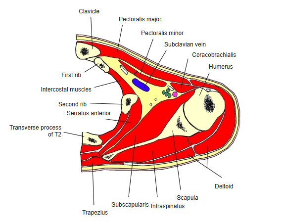
Origin / insertion
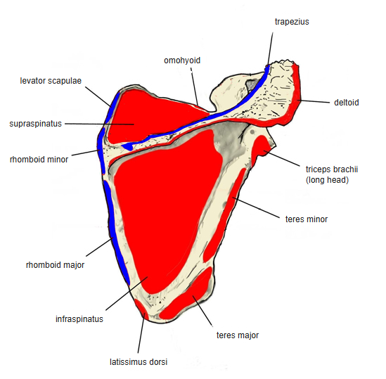
- Origin:
- Clavicular part: lateral third of the clavicle
- Acromial part: acromion of the scapula
- Spinal part: spine of the scapula
- Insertion: deltoid tuberosity of the humerus
- The fibres converge on the insertion: the middle ones run vertically, the anterior ones backward and the posterior ones forward.
Action
- Whole muscle: the main abductor of the arm
- Clavicular part: flexes and medially rotates the arm
- Acromial part: abducts the arm (some sources also list lateral rotation)
- Spinal part: extends and laterally rotates the arm
- Helps keep the head of the humerus in place when heavy loads are carried in the hand
Innervation
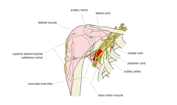
- Axillary nerve (C5–C6; some sources add C4)
- The axillary nerve can be injured in anterior dislocation of the shoulder and in operations in the axilla.
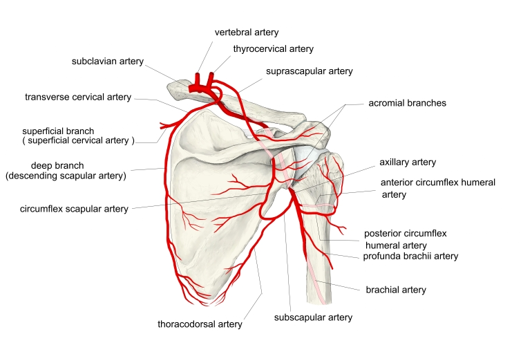
Blood supply
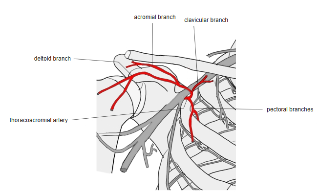
- Posterior circumflex humeral artery
- Thoracoacromial artery (acromial and deltoid branches)
- Deep artery of the arm (deltoid branch)
Related terms
- Muscles of the shoulder girdle
- Pectoralis major
- Trapezius
- Deltopectoral groove
- Deltopectoral triangle
- Cephalic vein
- Deltoid tuberosity
- Axillary nerve
- Posterior circumflex humeral artery
- Thoraco-acromial artery
- Deep artery of arm
References and sources
- Further reading: Deltoid muscle, Wikipedia.