Latin: nervus axillaris

Overview

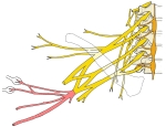
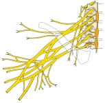
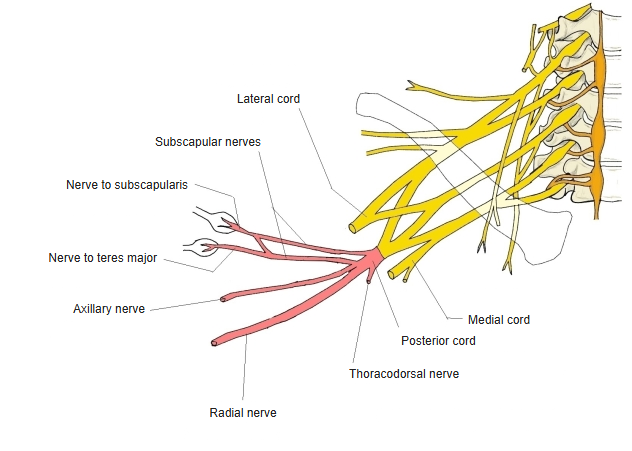
  • Schematic brachial plexus with the posterior cord highlighted in red: subscapular nerves, nerve to subscapularis, nerve to teres major, thoracodorsal, axillary and radial nerves; lateral and medial cords labelled
    Posterior cord and its branches
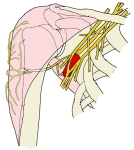
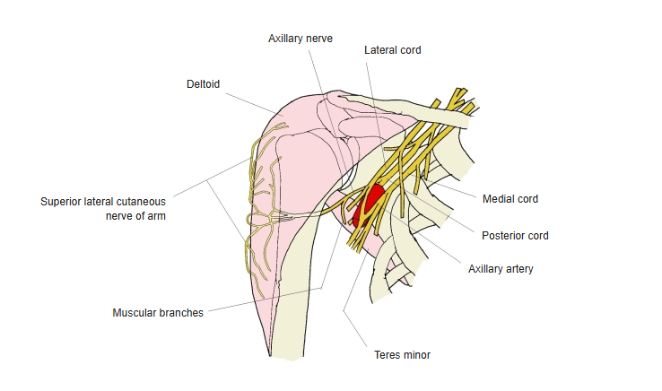
  • Right shoulder from the front showing the axillary nerve from the posterior cord, its muscular branches to deltoid and teres minor and the superior lateral cutaneous nerve of arm, with the axillary artery and the lateral, medial and posterior cords
    Axillary nerve (anterior view of the shoulder)
Branches

BranchSupply
Muscular branchesDeltoid, teres minor
Superior lateral cutaneous nerve of armSkin over the lower part of deltoid
Articular branchShoulder (glenohumeral) joint
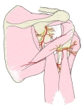
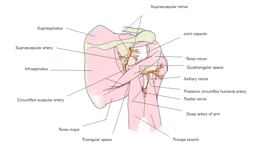
  • Posterior view of the right shoulder and arm showing the axillary nerve and posterior circumflex humeral artery in the quadrangular space, the circumflex scapular artery in the triangular space, the radial nerve and deep artery of arm, and surrounding muscles
    Quadrangular and triangular spaces (posterior view)
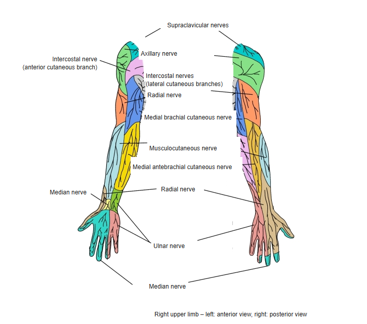
  • Front and back of the right upper limb with the skin areas of each cutaneous nerve in colour.
    Cutaneous nerves of the upper limb
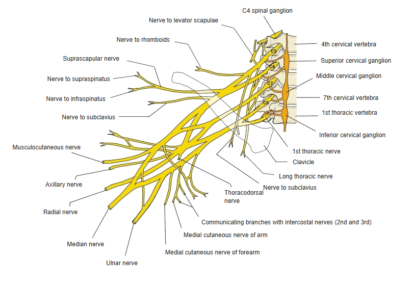
  • Schematic right brachial plexus from C5 to T1 with the cervical sympathetic ganglia, showing the supraclavicular branches and the terminal nerves of the upper limb
    Brachial plexus and its branches
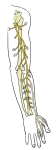
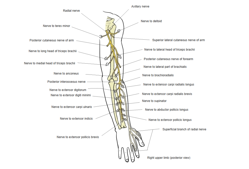
  • Right upper limb from behind showing the radial nerve with its muscular branches to triceps, anconeus, brachioradialis and the forearm extensors, its cutaneous branches, the posterior interosseous nerve and the superficial branch
    Radial nerve and its branches (posterior view)
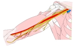
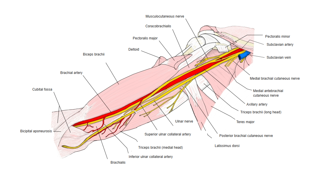
  • Medial view of the arm with the median, ulnar and musculocutaneous nerves, the brachial artery and nearby muscles.
    Nerves and vessels of the arm

References and sources

  1. Further reading: Axillary nerve, Wikipedia.