Latin: musculus gluteus maximus
Overview
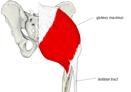
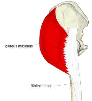
- The largest and most superficial of the gluteal muscles and the main extensor of the hip, especially important in walking.
- The iliopsoas in front of the pelvis flexes the hip; the gluteal muscles behind extend it.
- The muscle can be divided into superficial and deep parts.
- In standing its lower part covers the ischial tuberosity; when the thigh is flexed it moves up, so in sitting the tuberosity lies just under the skin and fat.
- The gluteal fold runs almost horizontally and deepens when the muscle contracts, but it does not mark the lower border of the muscle.
- Bursae:
- Trochanteric bursa: separates the tendon from the greater trochanter
- Ischial bursa: between the ischial tuberosity and the muscle; in people who sit on hard seats it can become inflamed and press on the posterior femoral cutaneous nerve
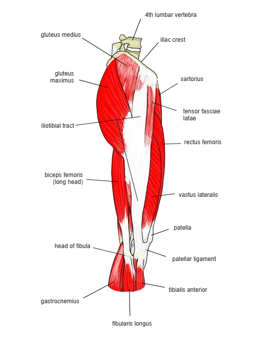
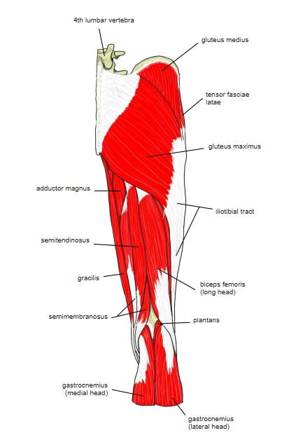
- Continuous in places with the gluteus medius, piriformis, quadratus femoris, vastus lateralis, vastus intermedius, tensor fasciae latae, adductor minimus and the short head of the biceps femoris.
Position in the classification
Lower Limb › Hip › Outer hip muscles › Gluteal muscles
- Gluteus maximus
- Gluteus medius
- Gluteus minimus
Illustrations
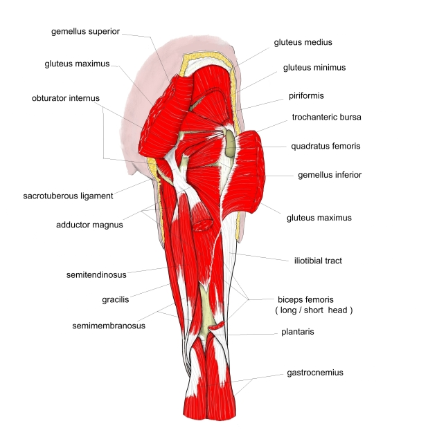
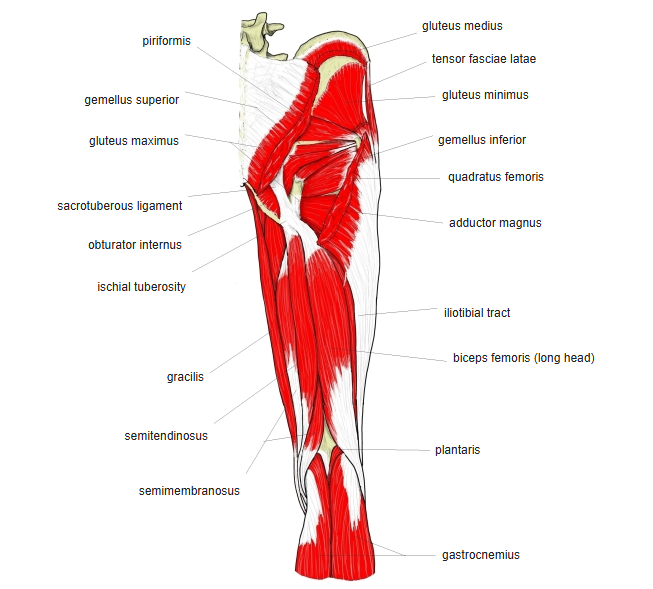
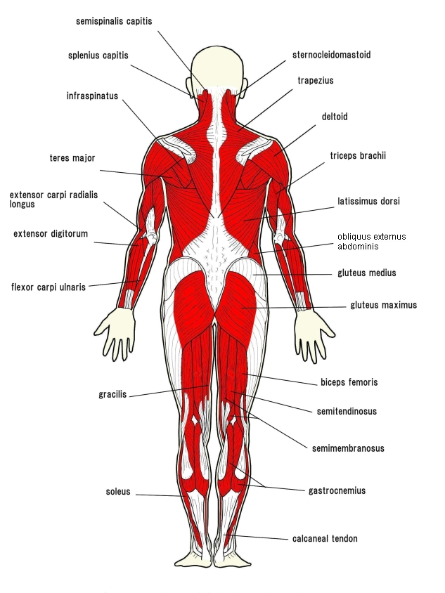
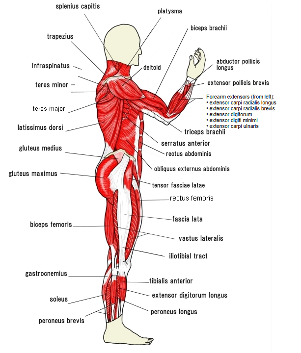
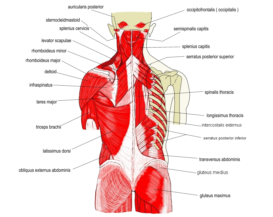
Origin / insertion
- Origin:
- Gluteal (outer) surface of the ala of the ilium behind the posterior gluteal line, and the iliac crest behind that line
- Lateral margins of the sacrum and coccyx
- Thoracolumbar fascia
- Sacrotuberous ligament
- Insertion:
- Superficial layer: iliotibial tract
- Deep layer: gluteal tuberosity of the femur (some sources also include the lateral lip of the linea aspera below it)
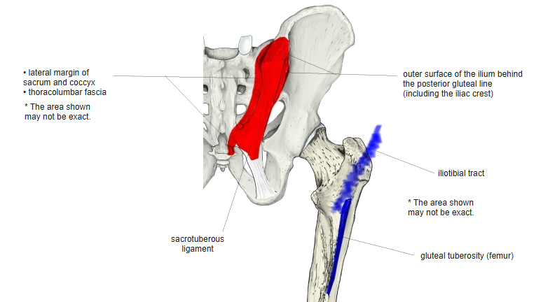
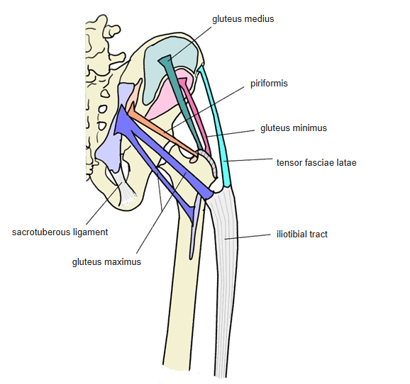
Action
- Extends the hip joint; also rotates the thigh laterally
Innervation
- Inferior gluteal nerve (L5–S2; some sources L4–S2)
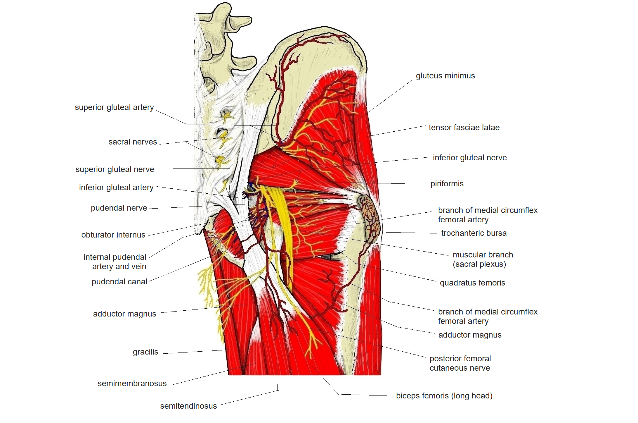
Blood supply
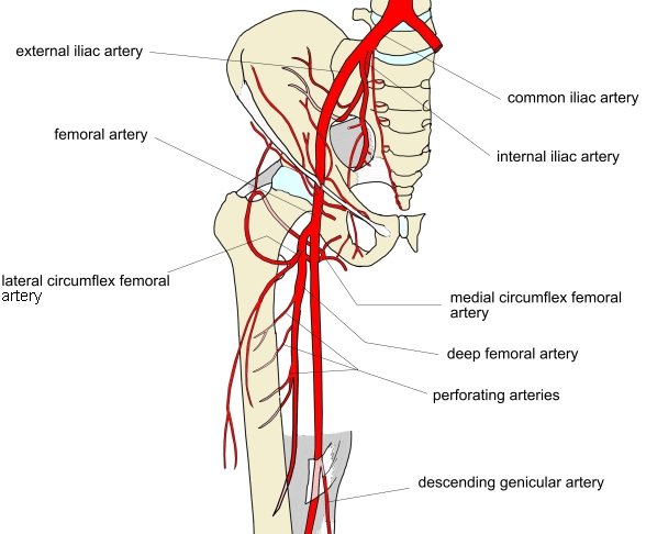
- Superior and inferior gluteal arteries, medial circumflex femoral artery, first perforating artery of the deep femoral artery
Related terms
- Gluteus medius
- Gluteus minimus
- Tensor fasciae latae
- Iliotibial tract
- Sacrotuberous ligament
- Gluteal tuberosity
- Inferior gluteal nerve
- Inferior gluteal artery
References and sources
- Further reading: Gluteus maximus, Wikipedia.