Latin: musculus gluteus minimus
Overview
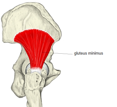
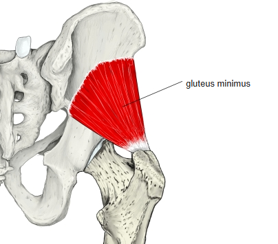
- The smallest of the three gluteal muscles; fan-shaped.
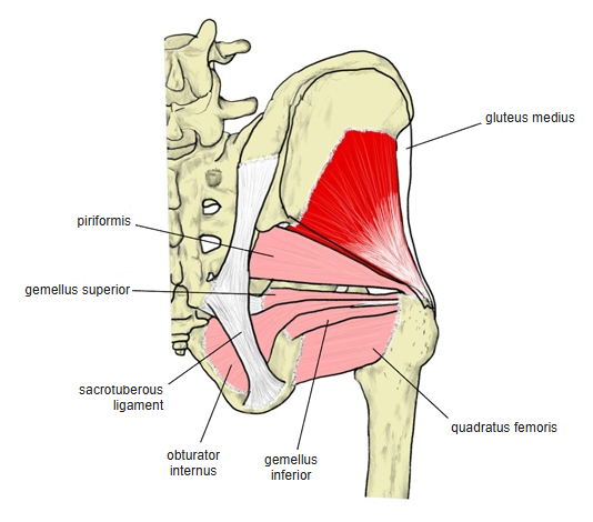
- Almost completely covered by the gluteus medius; the two muscles are fused along their anterior borders, develop from a common primordium, and together form the small gluteal muscles.
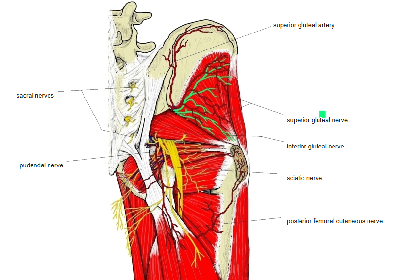
- The deep branches of the superior gluteal vessels and the superior gluteal nerve run between the gluteus medius and the gluteus minimus.
- A bursa (trochanteric bursa of the gluteus minimus) lies between its tendon and the greater trochanter.
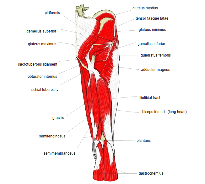
- Continuous in places with the piriformis, gluteus medius and tensor fasciae latae.
Position in the classification
Lower Limb › Hip › Outer hip muscles › Gluteal muscles
- Gluteus maximus
- Gluteus medius
- Gluteus minimus
Illustrations
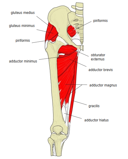
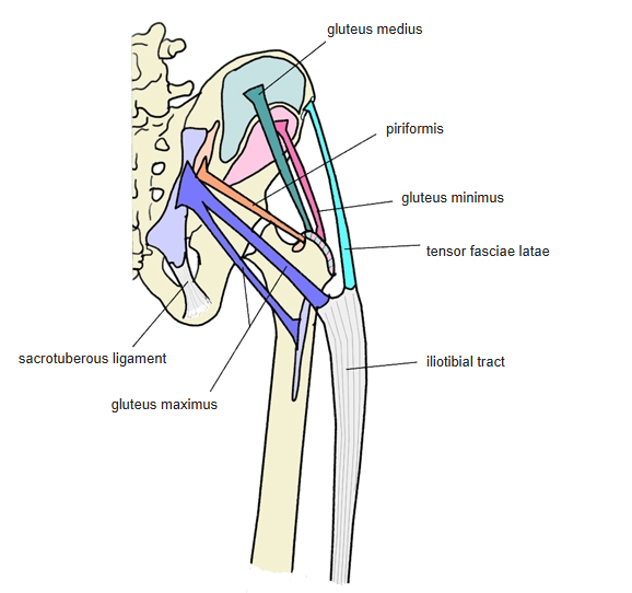
Origin / insertion
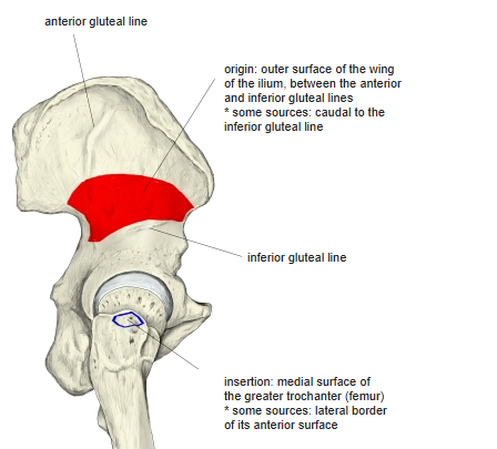
- Origin: outer surface of the ilium
- Between the anterior and inferior gluteal lines
- Also the area below the inferior gluteal line (some sources)
- Behind, also the margin of the greater sciatic notch
- Insertion: greater trochanter:
- Anterior surface (toward its lateral border)
- Also sends an expansion to the capsule of the hip joint
Action
- Abducts the hip
- With the gluteus medius, it keeps the pelvis level during single-leg stance and walking
- Anterior fibres help to flex and medially rotate the hip
- Weakness of the gluteus medius and minimus lets the pelvis drop on the opposite side (Trendelenburg sign); walking becomes a Duchenne gait, or a waddling gait when both sides are weak
Innervation
- Superior gluteal nerve (L4–S1)
Blood supply
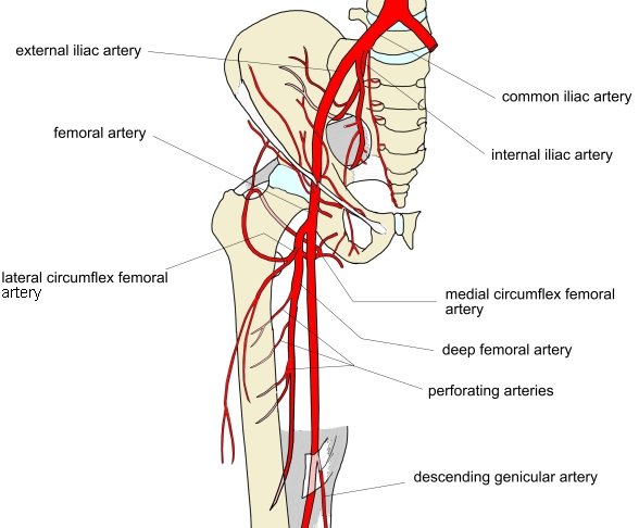
- Superior gluteal artery
- Lateral circumflex femoral artery
Related terms
- Gluteus medius
- Gluteus maximus
- Piriformis
- Tensor fasciae latae
- Ilium
- Greater trochanter
- Superior gluteal nerve
- Superior gluteal artery
- Lateral circumflex femoral artery
- Trendelenburg sign
References and sources
- Further reading: Gluteus minimus, Wikipedia.