Latin: musculus gluteus medius
Overview
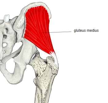
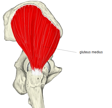
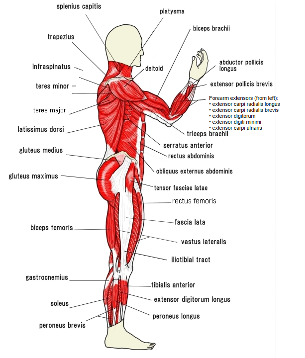
- A broad, thick, fan-shaped muscle on the outer surface of the pelvis; triangular when seen from the side.
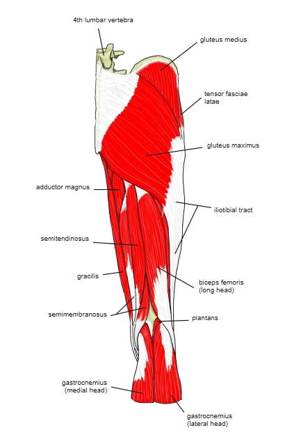
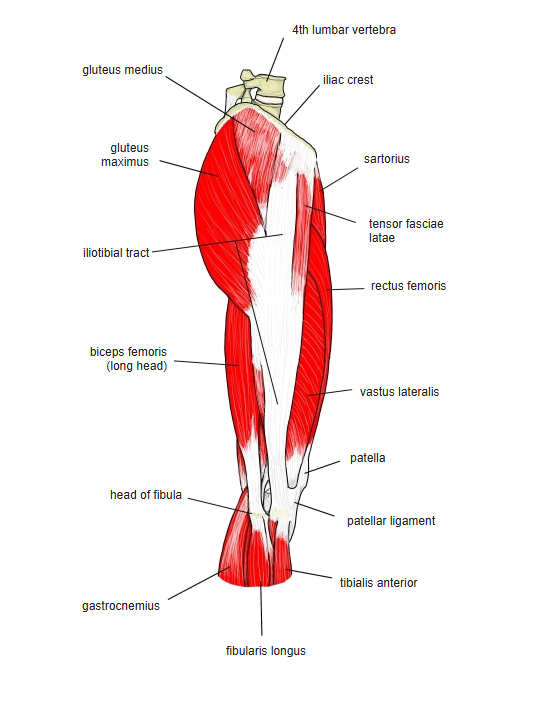
- Its anterior two-thirds are covered by the gluteal aponeurosis and its posterior third by the gluteus maximus.
- A bursa (trochanteric bursa of the gluteus medius) lies between it and the greater trochanter.
- With the gluteus minimus it steadies the pelvis in standing and walking. If these abductors are weak on one side, the pelvis drops on the opposite side when standing on that leg: Trendelenburg sign; walking shows a Duchenne limp, and weakness on both sides gives a waddling gait.
- Continuous in places with the gluteus maximus and minimus, tensor fasciae latae, piriformis and vastus lateralis.
Position in the classification
Lower Limb › Hip › Outer hip muscles › Gluteal muscles
- Gluteus maximus
- Gluteus medius
- Gluteus minimus
Illustrations
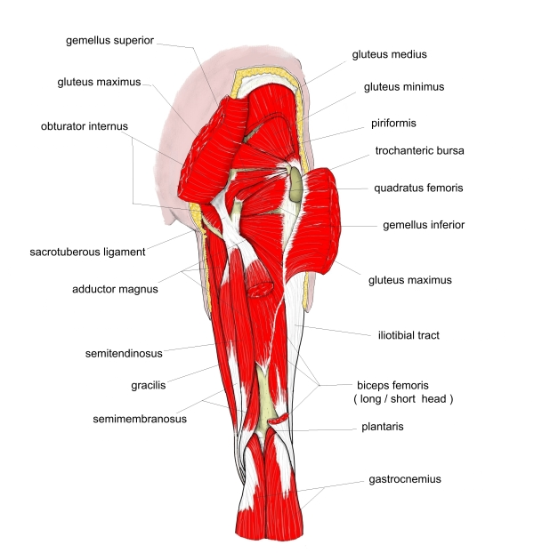
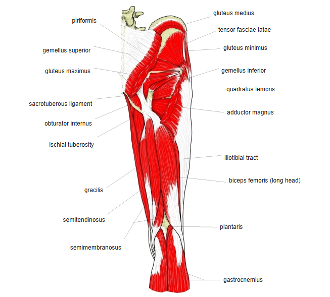
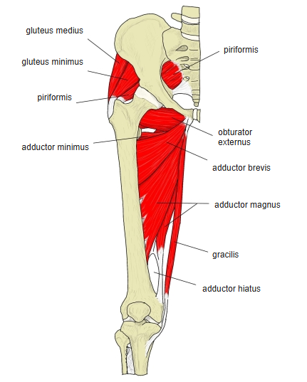
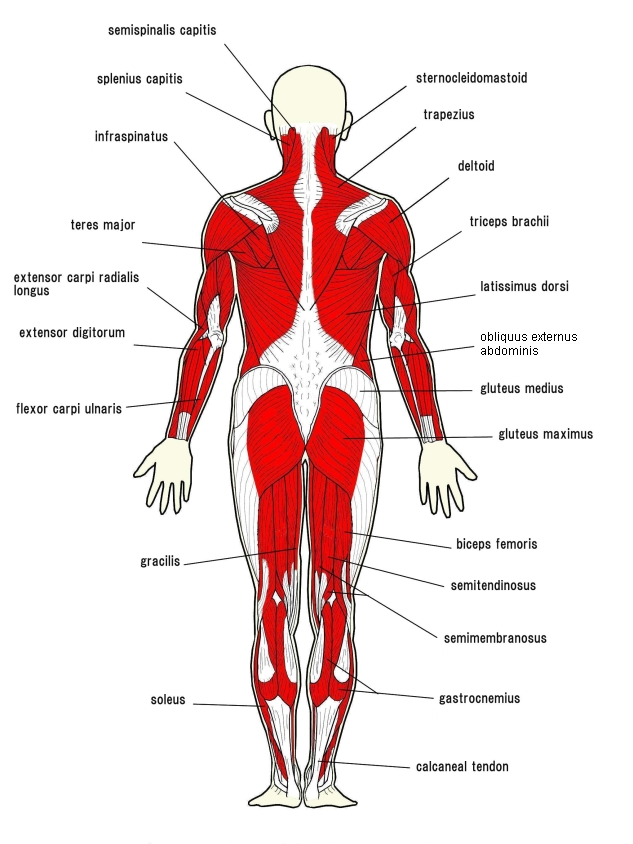
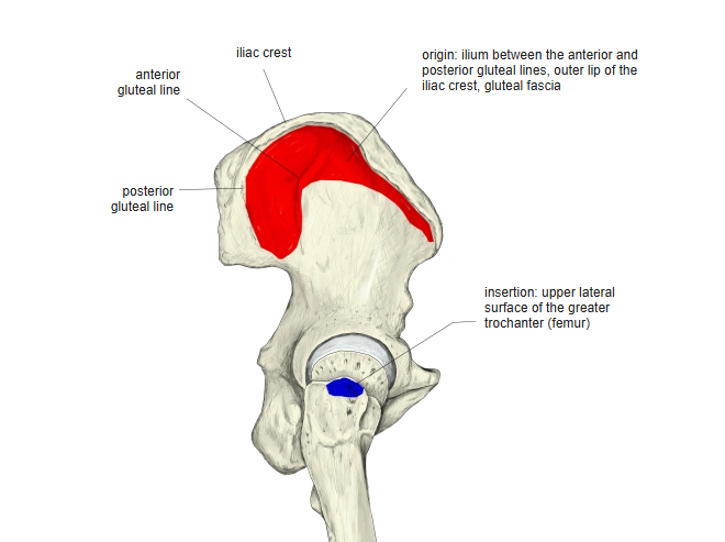
Origin / insertion
- Origin: outer surface of the ilium between the anterior and posterior gluteal lines, up to the iliac crest, and the gluteal aponeurosis
- Insertion: oblique ridge running downward and forward on the lateral surface of the greater trochanter
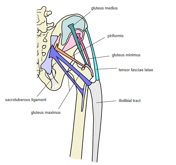
Action
- Abducts the hip and steadies the pelvis
- The anterior fibres help to flex and medially rotate the hip; the posterior fibres help to extend and laterally rotate it
Innervation
- Superior gluteal nerve (L4–S1; some sources L4–S2)
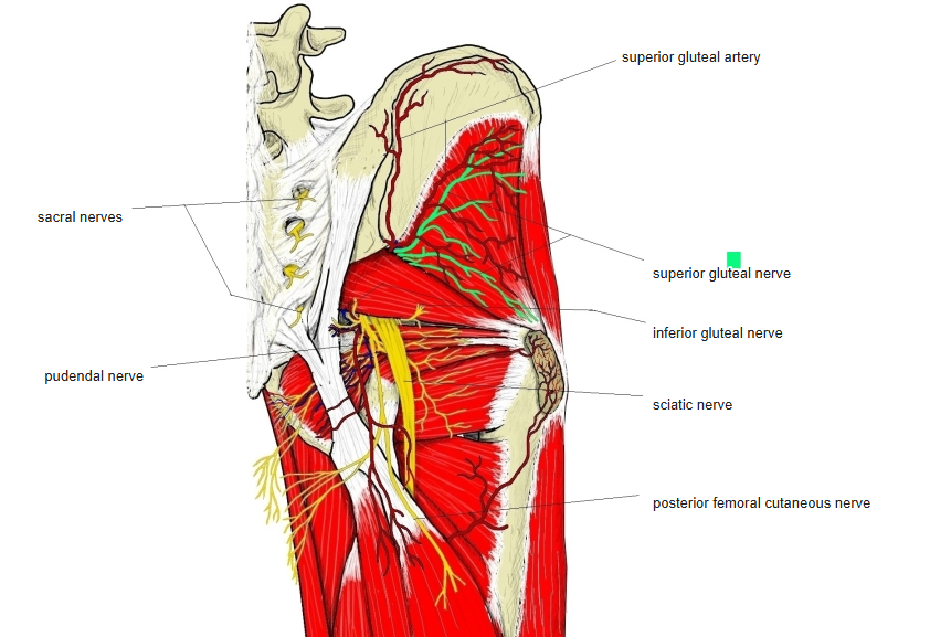
Blood supply
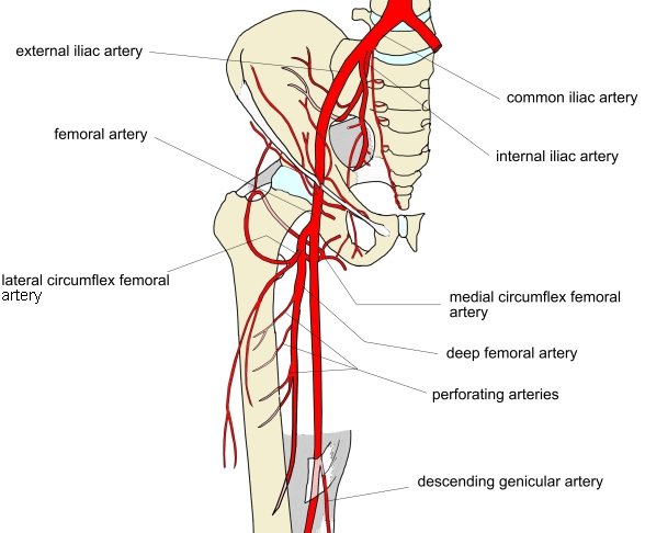
- Superior gluteal artery, lateral circumflex femoral artery
Anatomical variations
- The posterior border may be closely joined to the piriformis, or some fibres end on its tendon
Related terms
- Gluteus maximus
- Gluteus minimus
- Tensor fasciae latae
- Piriformis
- Greater trochanter
- Superior gluteal nerve
References and sources
- Further reading: Gluteus medius, Wikipedia.