Latin: musculus levator scapulae
Overview
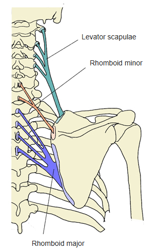
- One of the superficial back muscles, in the second layer with the rhomboid major and minor.
- As its name says, it raises the scapula.
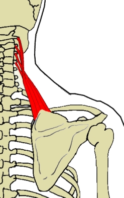
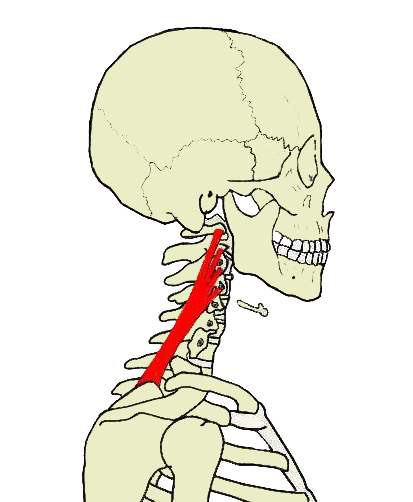
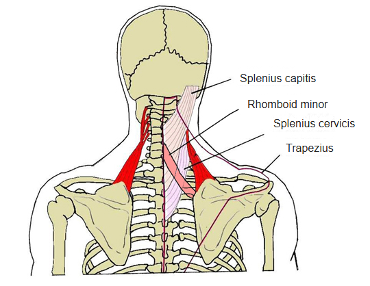
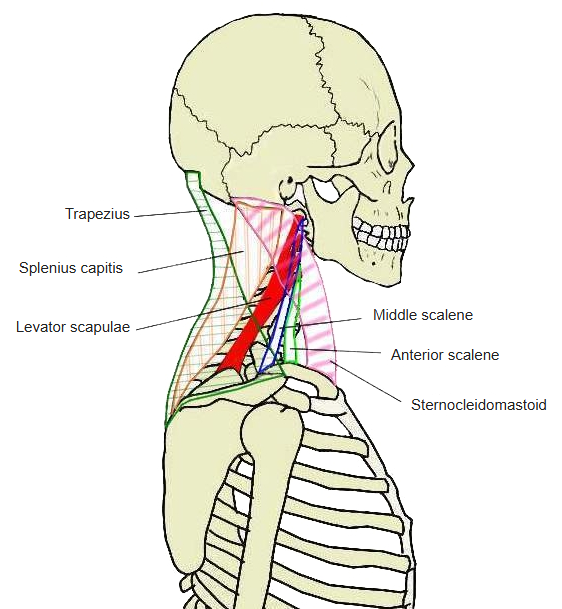
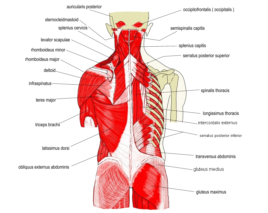
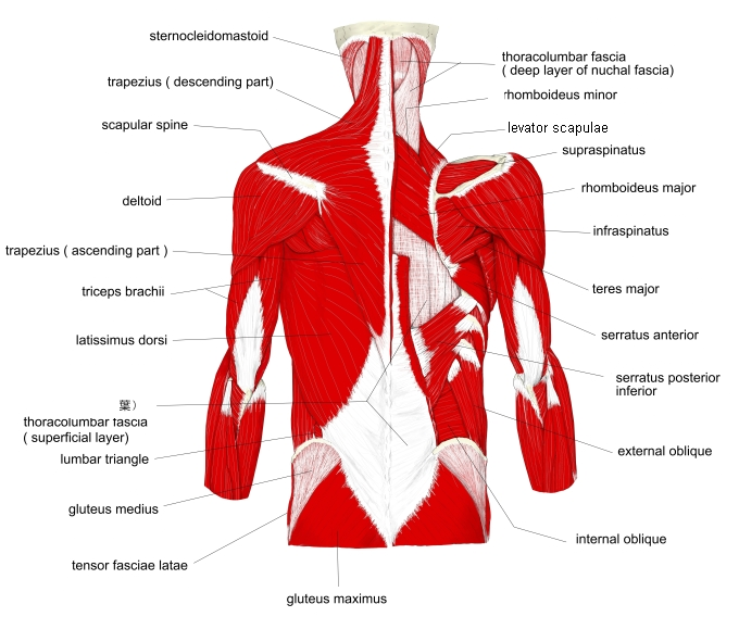
- Seen from behind it lies under the trapezius, joining the cervical vertebrae to the scapula in four or five muscular slips.
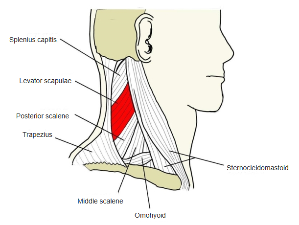
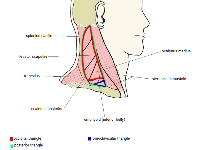
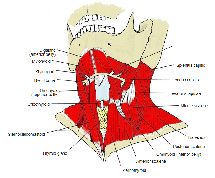
- Forms part of the floor of the posterior triangle of the neck, where part of it lies just under the skin.
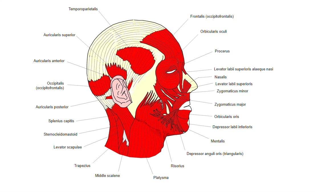
- Upper part covered by the sternocleidomastoid, lower part by the trapezius; the middle scalene lies in front and the splenius cervicis behind.
Position in the classification
Trunk › Back › Superficial back muscles › Second layer
- Rhomboid major
- Rhomboid minor
- Levator scapulae
Illustrations
Origin / insertion
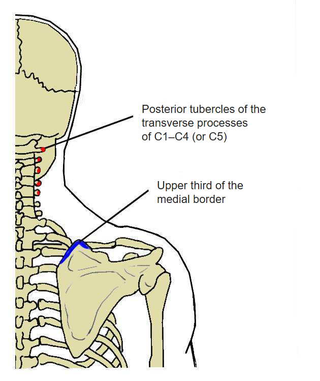
- Origin: transverse processes of the atlas and axis (C1–C2) and the posterior tubercles of the transverse processes of C3–C4 (sometimes C5)
- Insertion: medial border of the scapula between the superior angle and the root of the spine
Action
- With the neck fixed: raises the superior (medial) angle of the scapula and turns its inferior angle medially (with the rhomboids and pectoralis minor, downward rotation of the scapula)
- With the shoulder fixed: bends the neck to the same side and turns it to the same side
Innervation
- Third and fourth cervical nerves (C3–C4), and often a branch of the dorsal scapular nerve (C5)
- The accessory nerve crosses the middle of the muscle on its superficial surface; the dorsal scapular nerve lies deep to it or pierces it.
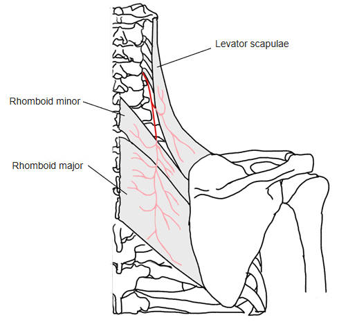
Blood supply
- Dorsal scapular artery (descending scapular artery)
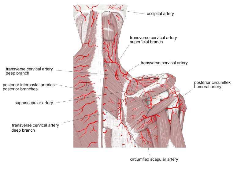
Anatomical variations
- The number of slips varies; a slip may reach the occipital bone or mastoid process, the trapezius, the scalenes, the serratus anterior, or the 1st or 2nd rib
- May be divided into several separate parts from origin to insertion
- Levator claviculae: from the transverse processes of one or two upper cervical vertebrae to the lateral end of the clavicle (corresponds to a muscle of other mammals)
- Often more or less united with the serratus anterior
Related terms
- Rhomboid major
- Rhomboid minor
- Trapezius
- Splenius cervicis
- Posterior triangle of the neck
- Dorsal scapular nerve
- Dorsal scapular artery
References and sources
- Further reading: Levator scapulae muscle, Wikipedia.
- Gray H. Anatomy of the Human Body, 20th ed. (1918), “7a. The Muscles Connecting the Upper Extremity to the Vertebral Column”, Bartleby.com.