Latin: musculus rectus femoris
Overview
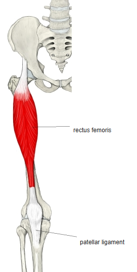
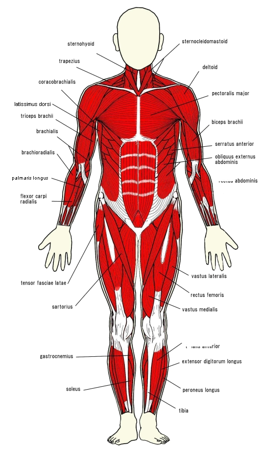
- The only two-joint muscle of the quadriceps femoris, in the middle of the front of the thigh.
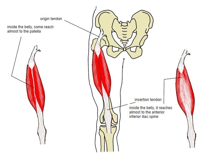
- Fusiform as a whole, but looks pennate from the surface (bipennate superficial fibres).
- Both tendons continue inside the belly: some fibres of the origin tendon reach almost to the patella, and the insertion tendon reaches almost to the anterior inferior iliac spine.
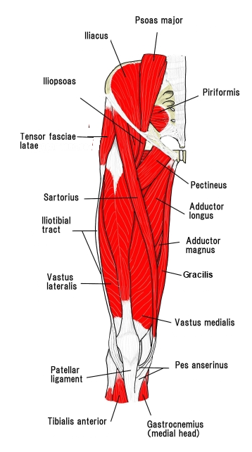
- Continuous in places with the iliacus, sartorius and the three vasti.
Position in the classification
Lower Limb › Thigh › Extensor group › Quadriceps femoris
- Rectus femoris
- Vastus medialis
- Vastus lateralis
- Vastus intermedius
Illustrations
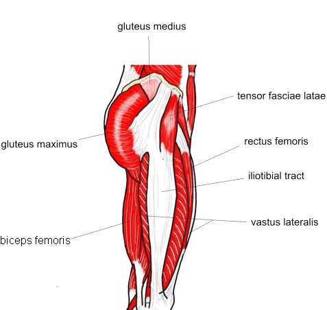
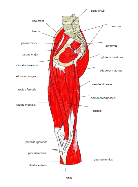
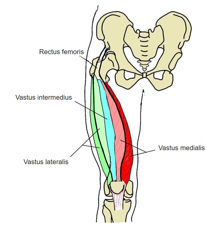
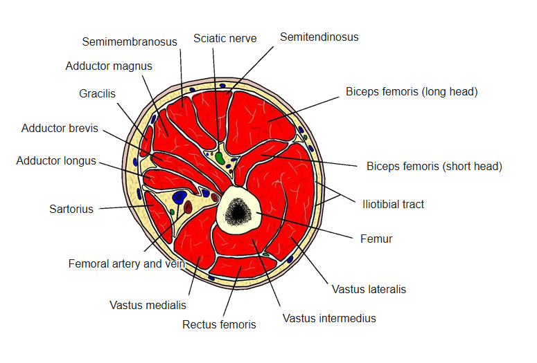
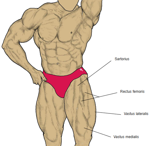
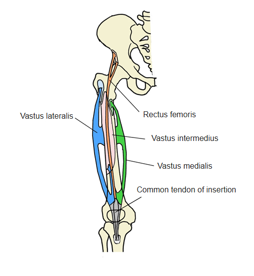
Origin / insertion
- Origin: anterior inferior iliac spine and the upper rim of the acetabulum (ilium)
- Insertion: base of the patella and, partly by way of the patellar ligament, the tibial tuberosity; many sources give only the patella
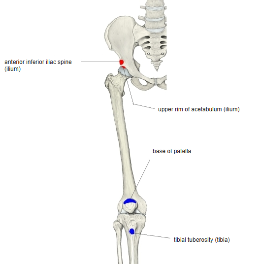
Action
- Raises the thigh (flexes the hip); in standing, flexes the pelvis
- As a two-joint muscle it also extends the knee
Innervation
- Femoral nerve (L2–L4)
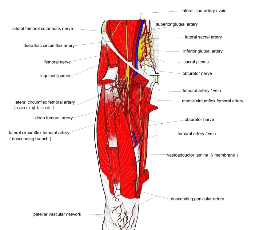
Blood supply
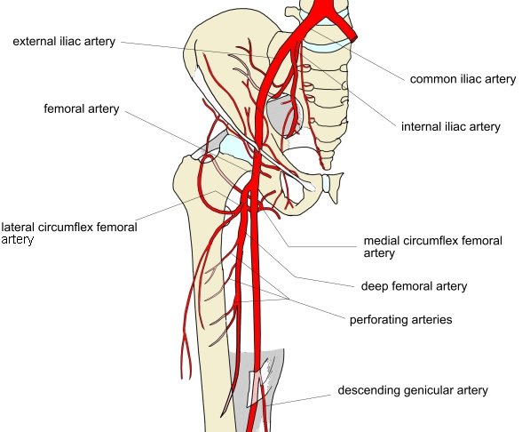
- Lateral circumflex femoral artery
Related terms
- Vastus lateralis
- Vastus medialis
- Vastus intermedius
- Quadriceps femoris
- Anterior inferior iliac spine
- Patella
- Tibial tuberosity
- Femoral nerve
References and sources
- Further reading: Rectus femoris muscle, Wikipedia.