Latin: musculus vastus intermedius
Overview
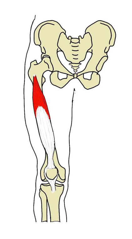
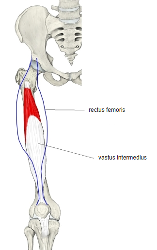
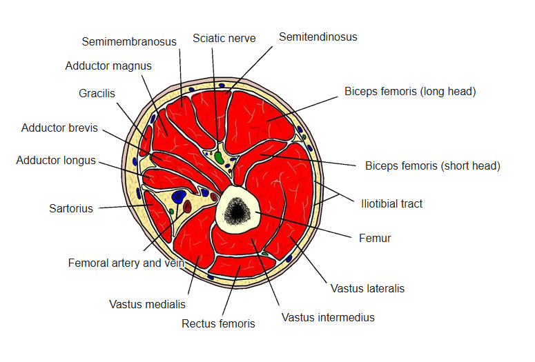
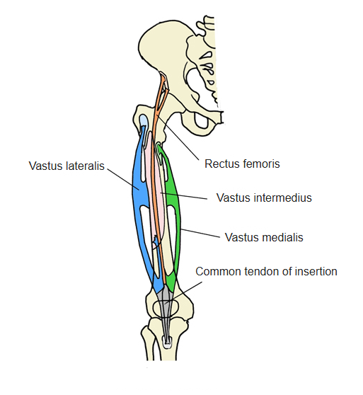
- The deepest part of the quadriceps femoris, lying under the rectus femoris and covered on both sides by the vastus medialis and lateralis.
- The rectus femoris glides over the aponeurotic tendon on its front.
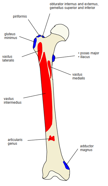
- Its most distal fibres end on the knee joint capsule as the articularis genus, which stops the capsule being pinched when the knee extends.
- Often hard to separate from the vastus medialis and lateralis, because the common tendon is split into 2–3 layers.
- Illustrations differ: fibres running straight or pennate, and more or less of the muscle drawn as tendon.
- Continuous in places with the other quadriceps muscles, adductor magnus, gluteus maximus and short head of the biceps femoris.
Position in the classification
Lower Limb › Thigh › Extensor group › Quadriceps femoris
- Rectus femoris
- Vastus medialis
- Vastus lateralis
- Vastus intermedius
Illustrations
Origin / insertion
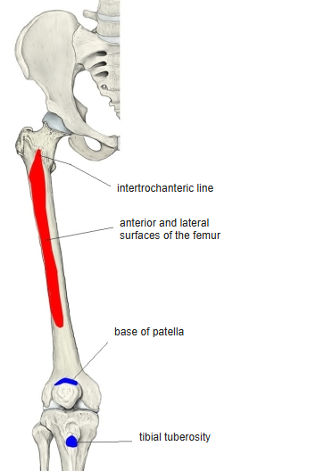
- Origin: front of the femur; some sources: front and lateral surfaces, from the intertrochanteric line to the distal two-thirds of the shaft
- Insertion: base of the patella and, partly by way of the patellar ligament, the tibial tuberosity
Action
- Extends the knee; tenses the medial patellar retinaculum
Innervation
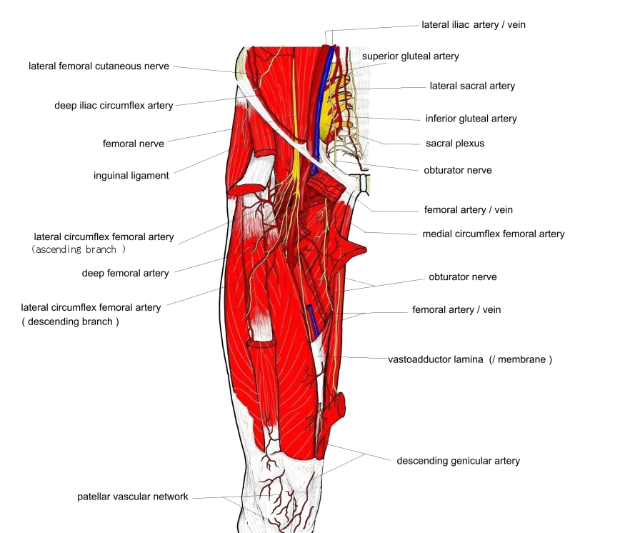
- Femoral nerve (L2–L4)
Blood supply
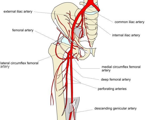
- Lateral circumflex femoral artery
Related terms
- Rectus femoris
- Vastus lateralis
- Vastus medialis
- Articularis genus
- Quadriceps femoris
- Patella
- Femoral nerve
References and sources
- Further reading: Vastus intermedius muscle, Wikipedia.