Latin: musculus temporoparietalis
Overview
- A thin sheet of muscle over the temporal region, very variable in extent and thickness (Rauber-Kopsch).
- The temporalis lies deep to it.
- The frontal belly is said to have separated from it; found in about 56% of people in one study.
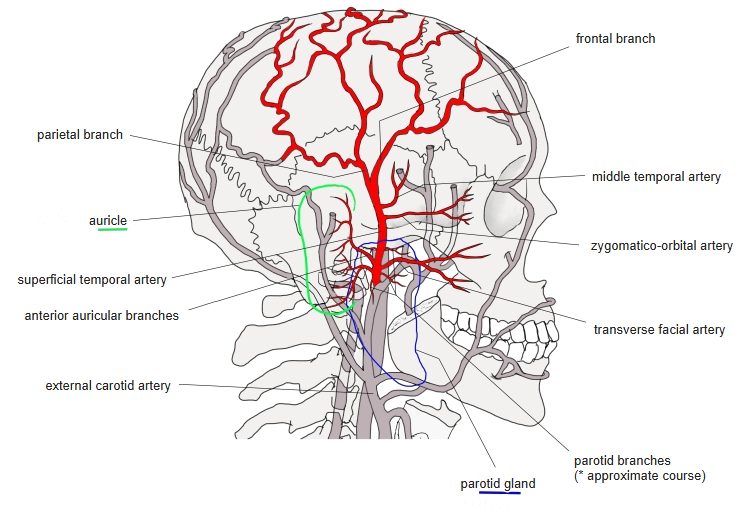
- Divided into two or three parts by the superficial temporal artery.
- Sometimes grouped with the occipitofrontalis as the epicranius.
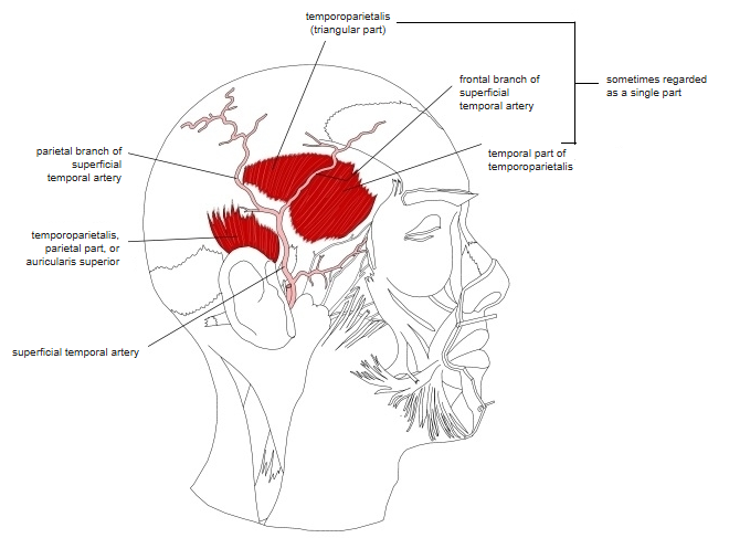
Position in the classification
Head › Facial muscles › Epicranius
- Frontalis
- Occipitalis
- Temporoparietalis
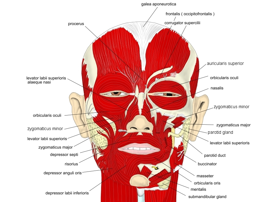
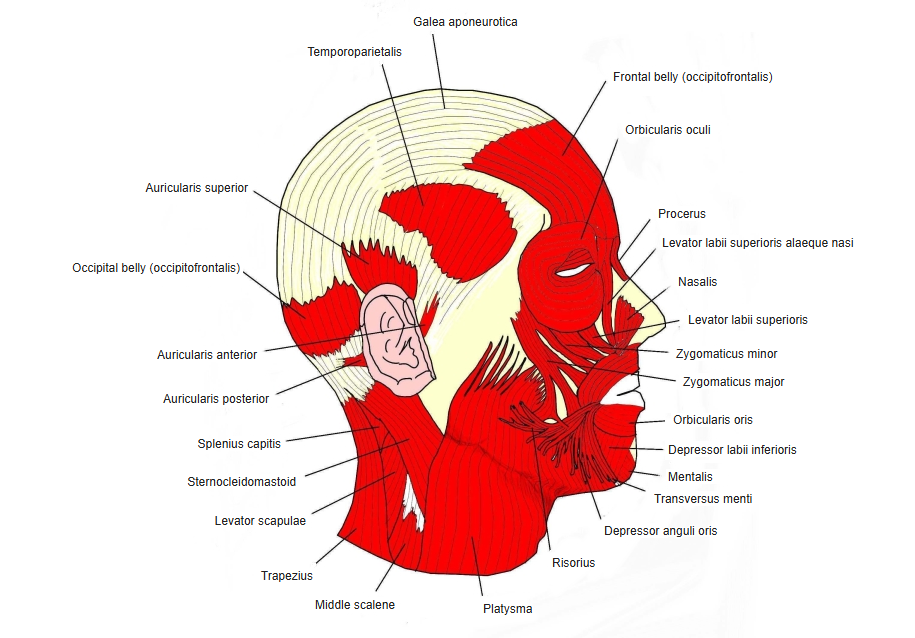
Illustrations
Origin / insertion
- Origin: lateral edge of the central part of the epicranial aponeurosis
- Insertion: upper part of the medial surface of the auricular cartilage
Action
- Pulls the auricle upwards
- May help the frontal belly to draw the epicranial aponeurosis forwards (Wikipedia)
Innervation
- Temporal branches of the facial nerve and the posterior auricular nerve (a branch of the facial nerve)
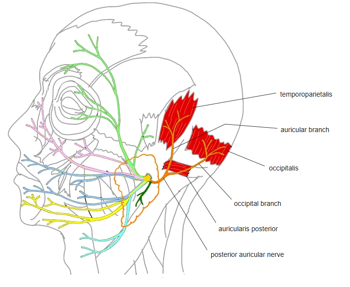
Blood supply
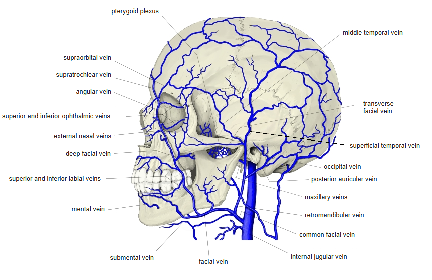
- Probably the superficial temporal artery and vein, which divide the muscle, but no source mentions them
Related terms
- Temporalis
- Frontal belly (occipitofrontalis)
- Auricularis superior
- Epicranial aponeurosis
- Facial nerve
- Superficial temporal artery
References and sources
- Further reading: Temporoparietalis muscle, Wikipedia.