Latin: nervus petrosus minor
Overview
- Carries parasympathetic secretory fibres for the parotid gland from the glossopharyngeal nerve.
- The continuation of the tympanic nerve beyond the tympanic plexus.
- Ends in the otic ganglion; postganglionic fibres reach the parotid gland via the auriculotemporal nerve.
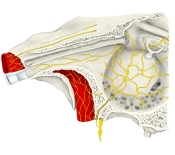
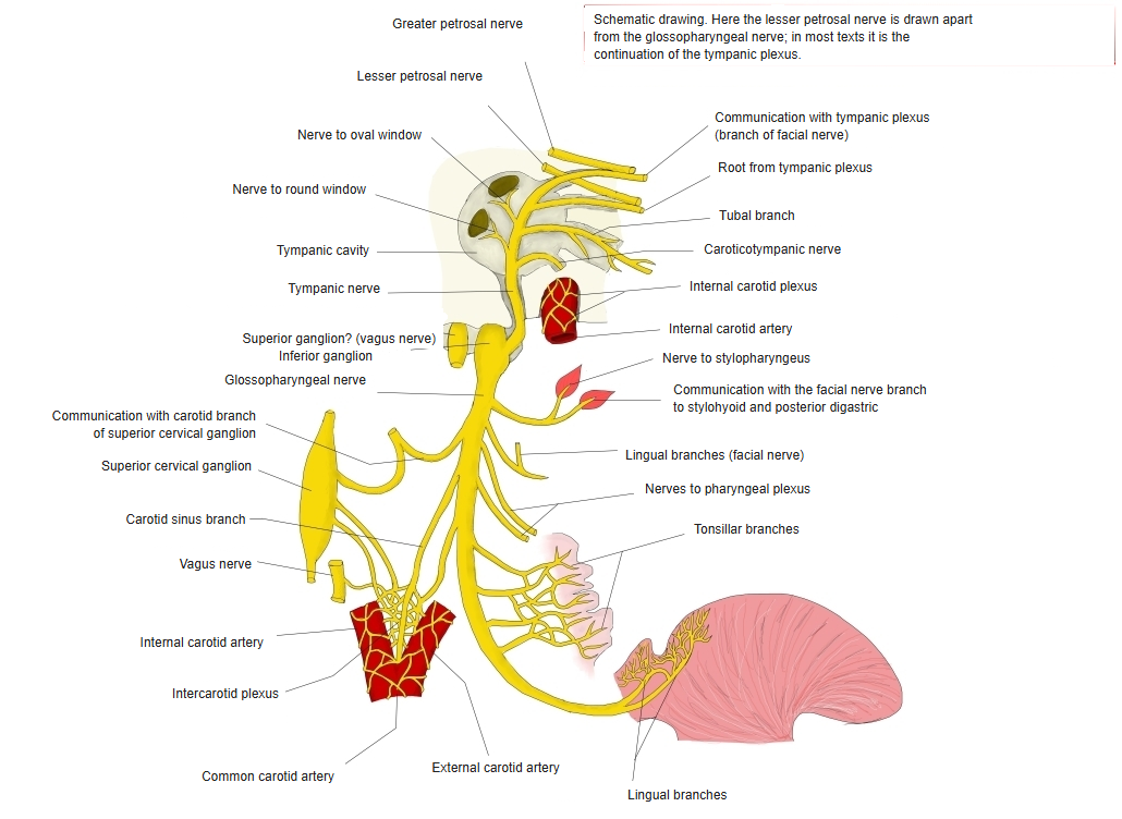
Tympanic plexus and glossopharyngeal nerve
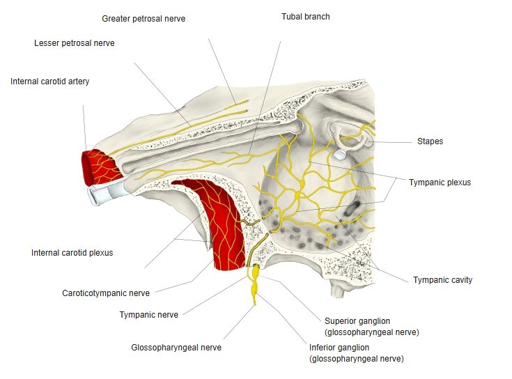
Course
- Leaves the tympanic cavity through the tympanic canaliculus.
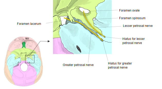
- Emerges on the anterior surface of the petrous part of the temporal bone at the hiatus for the lesser petrosal nerve and runs in its groove in the middle cranial fossa, under the dura.
- Passes through the foramen ovale (or the sphenopetrosal fissure) to the infratemporal fossa and enters the otic ganglion.
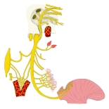
Glossopharyngeal nerve and its branches (schematic) 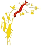
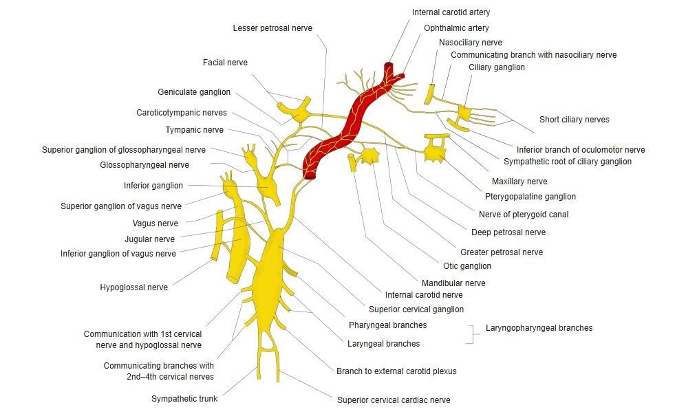
Cervical sympathetic trunk and the head (schematic) 
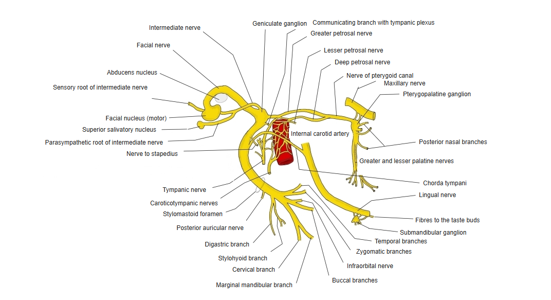
Facial nerve: nuclei and branches 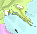
Hiatuses for the greater and lesser petrosal nerves
References and sources
- Further reading: Lesser petrosal nerve, Wikipedia.