Latin: nervus ophthalmicus
Overview
- The first and smallest division (V1) of the trigeminal nerve; purely sensory.
- Leaves the trigeminal ganglion, runs forwards in the lateral wall of the cavernous sinus and enters the orbit through the superior orbital fissure.
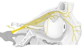
Ophthalmic nerve in the orbit (lateral view) 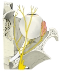
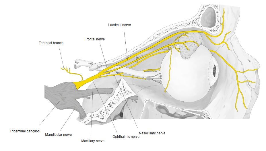
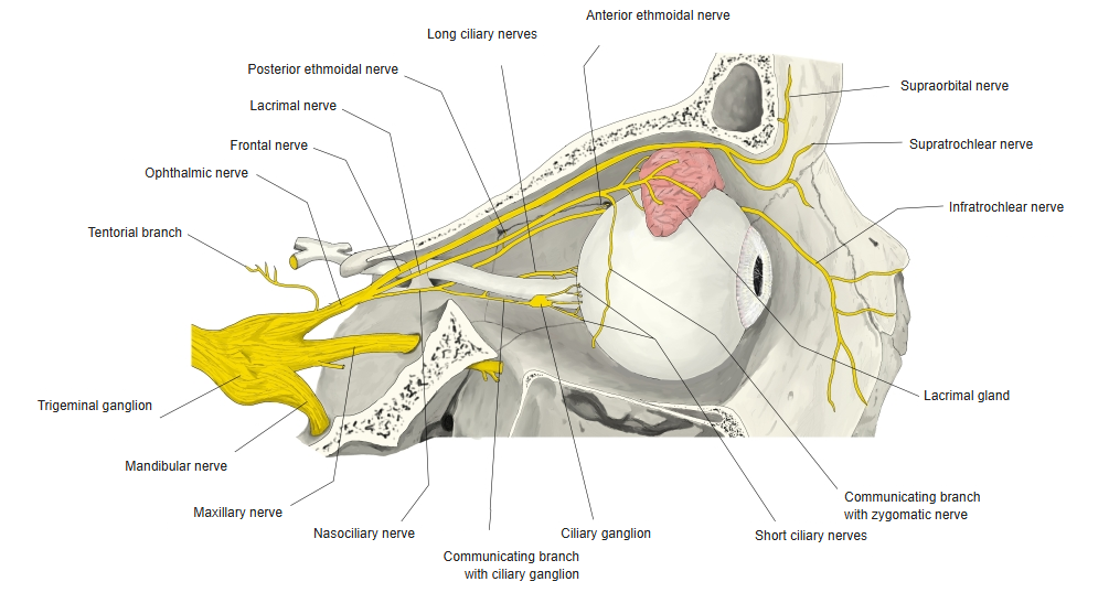
Branches of the ophthalmic nerve
Branches
- It communicates with the internal carotid plexus and with the oculomotor, trochlear and abducens nerves.
| Branch | Supply |
|---|---|
| Tentorial branch (inside the skull) | Tentorium cerebelli and nearby dura |
| Lacrimal nerve | Lacrimal gland, conjunctiva and skin of the lateral upper eyelid |
| Frontal nerve | Forehead and scalp via the supraorbital and supratrochlear nerves |
| Nasociliary nerve | Eyeball, ethmoidal air cells, nasal cavity and skin of the nose |
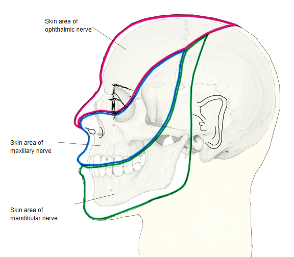
Skin areas of the trigeminal nerve
References and sources
- Further reading: Ophthalmic nerve, Wikipedia.