Latin: nervus ethmoidalis posterior
Overview
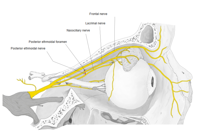
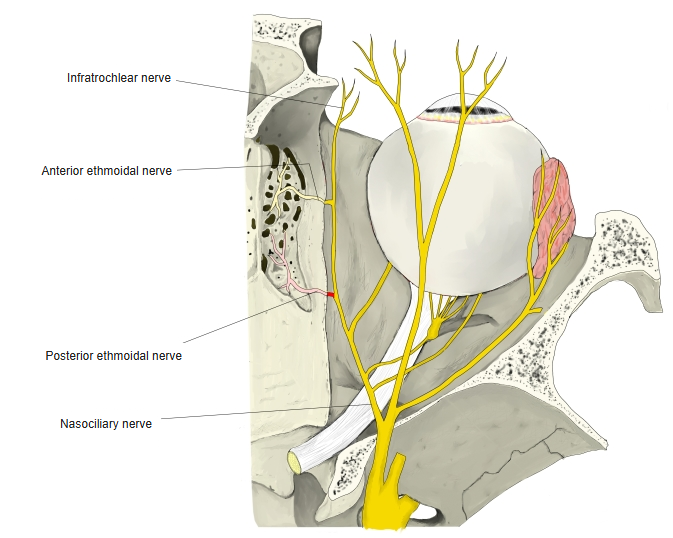
- A branch of the nasociliary nerve (ophthalmic division).
- Leaves near the posterior ethmoidal foramen and passes through it with the posterior ethmoidal artery.
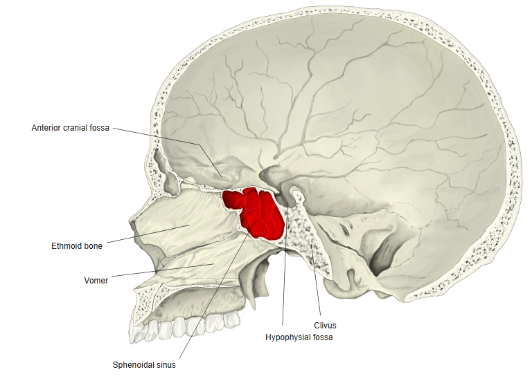
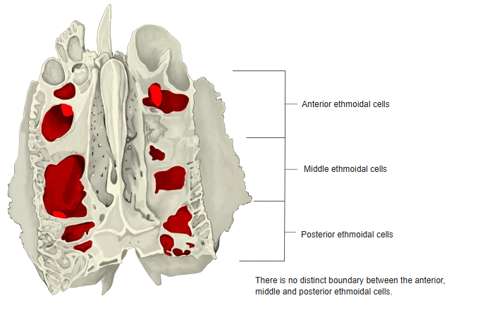
- Supplies the sphenoidal sinus and the posterior ethmoidal air cells; it is absent in about 30% of people.
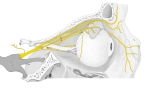
Posterior ethmoidal nerve (lateral view) 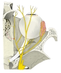
Ethmoidal nerves (medial wall of the orbit)
Area supplied
- Mucosa of the sphenoidal sinus and posterior ethmoidal cells.
Skull (sagittal section) 
Ethmoidal cells
References and sources
- Further reading: Posterior ethmoidal nerve, Wikipedia.