Latin: tunica conjunctiva
Overview
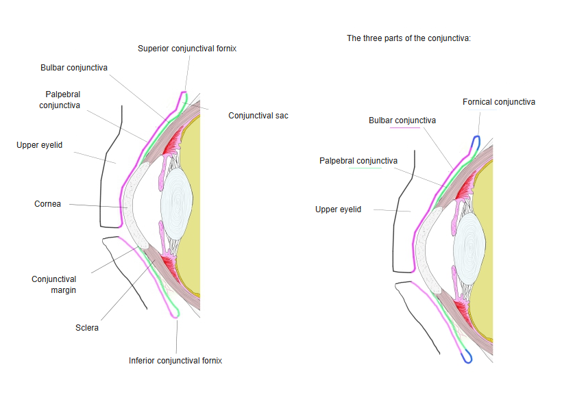
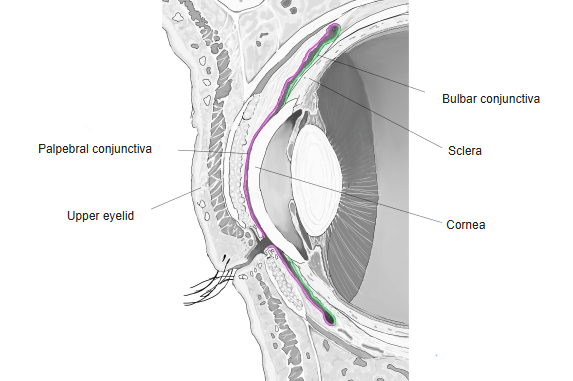
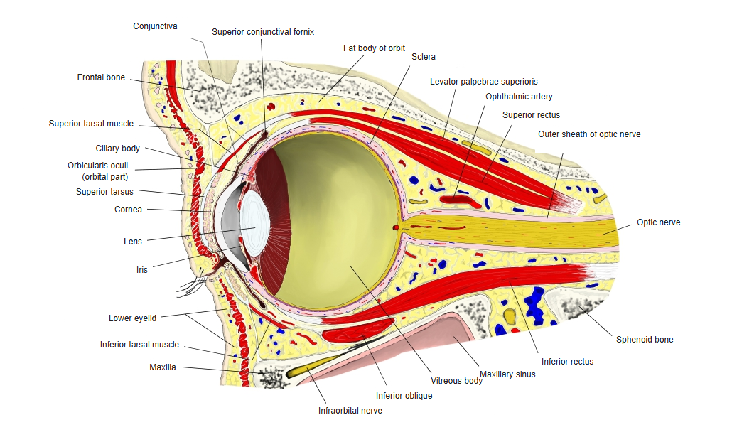
- A thin, transparent mucous membrane that lines the back of the eyelids and covers the front of the sclera up to the edge of the cornea.
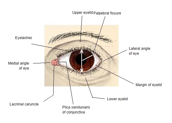
- The space it encloses between the eyelids and the eyeball is the conjunctival sac, open at the palpebral fissure.
- Its epithelium (stratified, with goblet cells) helps to keep the eye moist; small accessory lacrimal glands lie in it.
- Rich in blood vessels: they become visible as redness when it is inflamed (conjunctivitis).
Parts of the conjunctiva 
Conjunctiva, sagittal section
Parts
| Part | Features |
|---|---|
| Palpebral conjunctiva | Lines the back of the eyelids; firmly attached to the tarsus |
| Bulbar conjunctiva | Covers the sclera loosely; at the corneal limbus it continues into the corneal epithelium |
| Fornical conjunctiva | Folds between the two, forming the superior and inferior conjunctival fornices; the lacrimal ducts open into the superior fornix |
| Plica semilunaris | Crescent-shaped fold at the medial angle of the eye, beside the lacrimal caruncle |
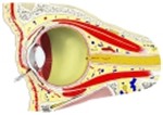
Orbit, sagittal section 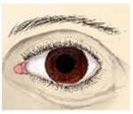
Eye, anterior view
Vessels and nerves
- Arteries: mainly the palpebral arches (palpebral and fornical parts) and, around the cornea, the anterior ciliary arteries; the two networks join.
- Nerves – upper part: supraorbital, supratrochlear and infratrochlear nerves.
- Lower part: infraorbital nerve. Lateral part: lacrimal nerve and the zygomaticofacial branch.
- Around the cornea: the long ciliary nerves.
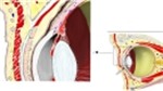
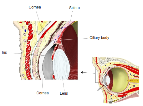
Front of the eyeball, sagittal section
References and sources
- Further reading: Conjunctiva, Wikipedia.