Latin: tunica fibrosa bulbi
Overview
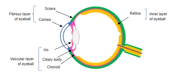
- The outer coat of the eyeball: tough, dense connective tissue that keeps the shape of the eye.
- Two parts: the opaque white sclera over the posterior five-sixths and the transparent cornea over the anterior one-sixth.
- The two meet at the corneal limbus, where the curvature changes and the scleral venous sinus drains the aqueous humor.
- Also called the fibrous tunic; it gives attachment to the extraocular muscles.
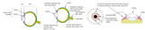
Sclera (diagram) 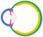
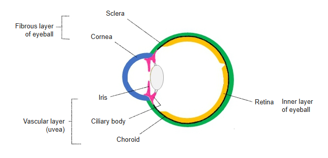
Three layers of the wall (diagram)
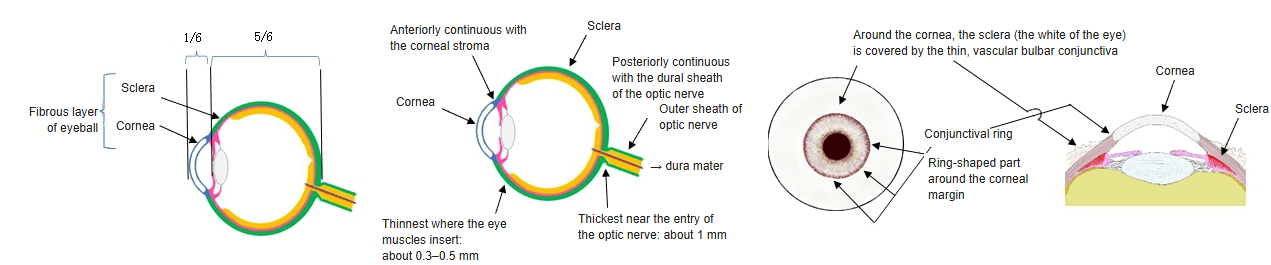
Sclera and cornea
| Sclera | Cornea | |
|---|---|---|
| Share of the coat | Posterior five-sixths | Anterior one-sixth |
| Appearance | Opaque, white ("white of the eye") | Transparent, more strongly curved |
| Collagen fibres | Irregular, interwoven | Regular layers of uniform fibrils |
| Blood vessels | Few | None |
| Continuous with | Dural sheath of the optic nerve (behind) | Conjunctival epithelium (at the limbus) |
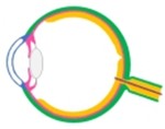
Layers of the eyeball wall (diagram) 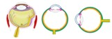
Retina and the layers of the eyeball wall
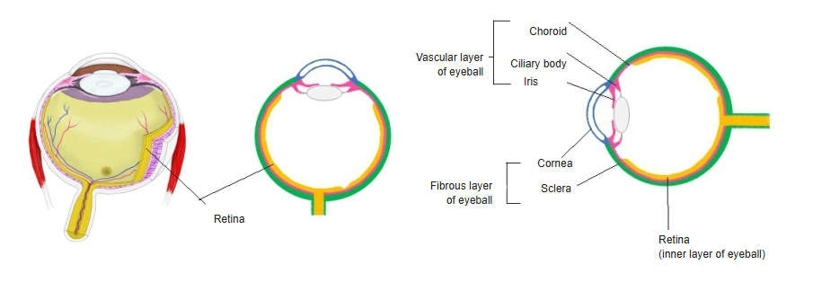
References and sources
- Further reading: Fibrous layer of eyeball, Wikipedia.